Looking for a student learning guide? It’s on the main menu for your course. Use the “Courses” menu above.
1. Introduction: Life is all about cell communication
For multicellular eukaryotes like us, every cell is a member of a team. And teamwork is all about communication.
Just think about what happens to you as you begin your day. After waking up, you decide to get out of bed. Cells in your brain send messages to your muscles, which respond by contracting in a coordinated series of movements that moves you from lying down to standing up. How do the cells in the brain communicate with the cells in your muscles?
Because it’s been a few hours since you’ve eaten, your blood sugar is a bit low. This condition is detected by cells in your pancreas. In response, these cells release a hormone called glucagon, which diffuses into your bloodstream, signaling the cells in your liver and skeletal muscles to break down the polysaccharide glycogen into glucose, which increases your blood sugar level.
As you eat breakfast, your blood sugar level rises. This condition is detected by a second set of cells in your pancreas, which secrete the hormone insulin into the bloodstream. When insulin is detected by cells in your liver and skeletal muscles, they open up glucose transport channels, allowing glucose to diffuse into the cells. Messages released inside the cells turn on enzymes that convert the glucose into glycogen, lowering your blood sugar level. Why do glucagon and insulin, which are secreted into the blood, only activate certain tissues? How do these hormones induce cells to change their activities?
Let’s see how cell communication works.
2. Cell Communication in Bacteria: Quorum sensing and Biofilms
The examples above focused on cell communication within our bodies. But cell communication isn’t limited to multicellular organisms, or even to eukaryotes. It also occurs in prokaryotes like bacteria.
An example of bacterial cell communication involves the creation of biofilms.
A biofilm is just what it sounds like: a living film (the word “film,” in this context, means thin layer). A biofilm is composed of a variety of microorganisms, primarily bacteria and fungi, that secrete an extracellular slime composed of polysaccharides, proteins, lipids, and DNA. Within the biofilm, these organisms support one another, and the biofilm creates optimal conditions for their growth.
While this is good for the living community within the biofilm, it can be bad for us. These biofilms can support pathogenic (disease-causing) bacteria.
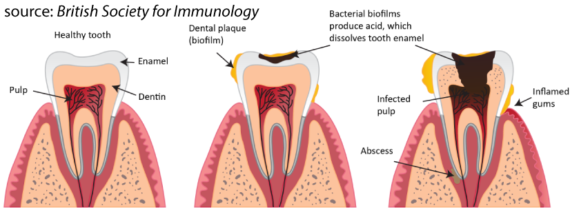
Plaque, for example, is the biofilm that forms on teeth. Bacteria in the plaque secrete acids that dissolve tooth enamel. As the enamel breaks down, the bacteria can infect the pulp (the tooth’s living tissue). They can also cause gum disease and break down the bone that holds the teeth in place.
How do these biofilms form?
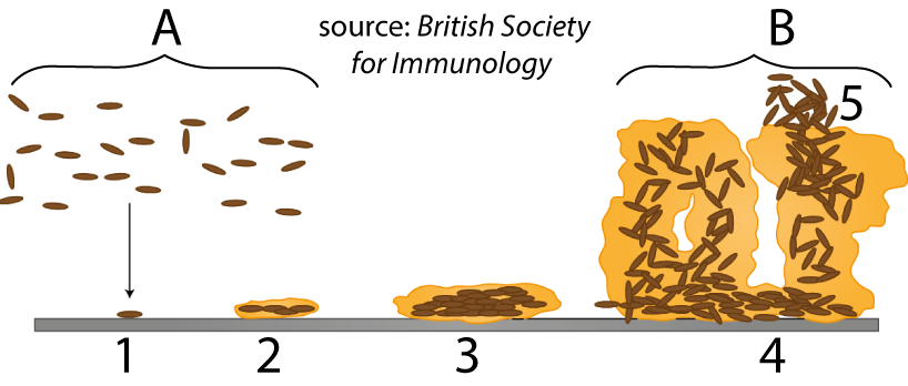
The bacteria in these biofilms also live in a free-floating independent form (at “A”). When they land on a surface (at “1”), they start to secrete the molecules that make up the molecular matrix of the biofilm. In “2,” “3,” and “4” you can see the biofilm growing into a three-dimensional structure, forming a colony. At a certain point, some of the bacteria within the colony detach (“5”) and revert to the free-floating form, enabling them to form new colonies on other surfaces.
Building a biofilm involves a type of cell communication called quorum sensing. A “quorum” is the minimum number of members that need to be present for an organization (anything from a student council to the U.S. Senate) to take a vote and make a binding decision. The basic idea behind quorum sensing is that the bacteria communicate with one another about their presence. At a certain density, the cells “decide” that the time has come to change their activities. In this case, the change is switching from the free-floating form to activating the genes that enable them to produce the biofilm.
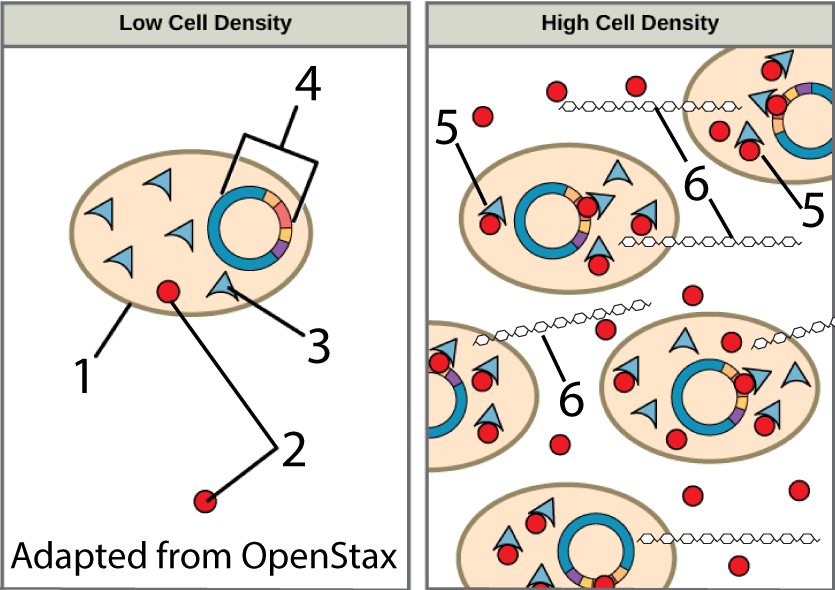
Here’s how this works. The bacterial cells (“1,” at left) produce signaling molecules (indicated by the red spheres at “2”). Signaling molecules are also called ligands: molecules that bind with a receptor (at “3”). When the bacterial cells are at a low density (left panel), the signaling molecules rarely bind with receptors, resulting in no cellular response.
As the density of the cells increases (right panel), so does the density of the ligands. The increased density results in more signals diffusing into the cells, where they bind with the receptors, forming a receptor-ligand complex (“5”). This complex binds with specific genes in the chromosomes of the bacterial cells (at “4”). Binding activates these genes, causing them to produce the polysaccharides (“6”) that comprise one component of the biofilm matrix.
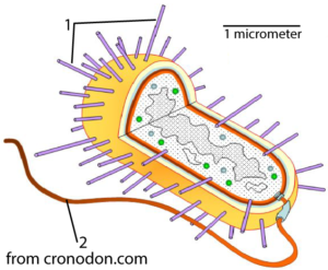
Quorum sensing also comes into play at the end of the process (step 5 in Figure 2, above). Bacteria can produce flagella (“2” in Figure 4 to your right). Flagella are little propellers embedded in the bacterium’s outer wall and membrane that give it mobility. In addition, the bacteria can produce pili, tiny rods that let them attach to a surface (at “1”). Bacteria use quorum sensing to determine when to switch from making flagella (which they use during their free-floating, mobile form), to making pili (which they use when they’re attaching to a surface, or locking themselves in place within a biofilm).
Quorum sensing illustrates some key elements of cell communication that we’ll see again and again throughout this unit. These include
- Release of a signal. The signal is a ligand: a molecule that binds to another molecule.
- Reception of the signal by a ligand-specific receptor.
- Induction of some cellular response. To induce means to persuade or influence someone to do something. Induction is the process of influencing, and it’s a term that’s used a lot in biology, particularly in developmental biology.
3. Cellular Communication in Yeast Mating
Let’s turn now to a unicellular eukaryote, and see how cell communication plays a role in one of life’s key processes: reproduction.
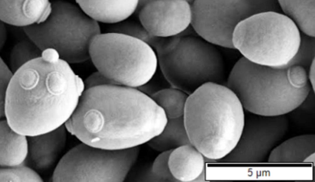
Saccharomyces cerevisiae, or Brewer’s Yeast, is a unicellular fungus that ferments sugar solutions into alcohol and carbon dioxide (creating the alcohol and bubbles in beer). You can buy Brewer’s Yeast for making beer or baking. Out of the package, it’s a granular, brownish powder (below left). Put it in some sugar water and look at it under a scanning electron microscope, and it looks like what’s shown below and to the right.
 |
 |
Yeast cells can exist in a haploid form with a single set of chromosomes, or a diploid form, with two sets of chromosomes. Both forms can reproduce asexually, by mitosis (replication of the entire set of chromosomes), followed by division into two daughter cells. The diploid form can also carry out a type of cell division called meiosis to produce the haploid form. That process parallels what happens when animals (including people) go through meiosis to create sperm cells and egg cells (which are also haploid). In the same way that meiosis in animals produces two types of sex cells — sperm and egg —meiosis in yeast results in two types of haploid cells: a-cells (pronounced “A cells”) and α-cells (“alpha cells”). And, just as fertilization in animals can only happen when a sperm cell fertilizes an egg cell, creating a diploid zygote, fertilization in yeast can only happen between an a-cell and α-cell (note that you’ll learn more about mitosis and meiosis later in this course).
You can follow this process in the diagram below. Start at “Y,” which shows a diploid yeast cell. This diploid cell resulted from the fusion of an a-cell and α-cell, which is why it’s labeled as “a/α.” The arrow at “Z” indicates meiosis, which produces the two haploid cells shown above the letter “V.”
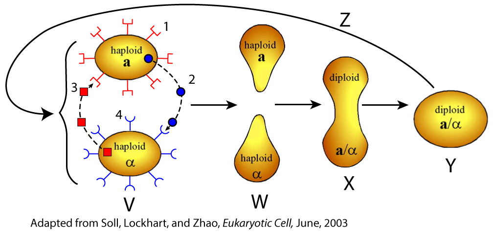
If we were examining meiosis in animals, then one of these haploid cells would be a sperm cell and the other would be an egg cell, and they’d have a correspondingly distinct appearance. The difference between the a-cell and α-cell is molecular. The a-cell has a receptor (at “1”) for the mating factor (at “3”) that’s produced by the α cell. The α cell has a receptor (at “4”) for the mating factor (at “2”) produced by the a-cell.
These mating factors are also called pheromones. Pheromones are chemical substances produced by one organism that affects the behavior or physiology of another organism. In this case, what these pheromones do is induce changes in the cell of the opposite mating type. When an a-cell receptor binds with pheromone released from an α-cell, the a-cell changes shape, becoming what’s called a “shmoo” (named for a comic book character from the 1940s). The α-cell does the same thing at the same time (see “W”), causing the two cells to grow toward one another. Eventually, they fuse (shown at “X”) and form a diploid zygote (at “Y”).
In terms of cell communication, the key things to focus on are
- The cells communicate through the production ligands. The ligands are the pheromones, or mating factors, shown at “2” and “3.”
- The ligands bind with specific receptors that have a shape that’s complementary to the ligand (shown at “1” and “4.”)
- The binding of the ligand with the receptor results in signal transduction that involves a second messenger. We’ll focus on this in the next tutorials. For now, all you need to know is that the initial signal (the mating factor) stops at the outside of the cell (where it binds with the receptor). Then, another signal is released that induces the cellular changes shown at “W.” It’s as if someone came knocking on your door, asking to speak to your roommate. The knock is the signal (and corresponds to the ligand). The door is the receptor. You answer the door, and then shout to your roommate, “Someone’s here for you.” You’re the second messenger, and your shout is signal transduction.
4. Checking Understanding: Quorum Sensing and Yeast Mating
[qwiz random = “true” qrecord_id=”sciencemusicvideosMeister1961-Cell Communication Overview 1 (quorum sensing, yeast mating)”] [h]
Cell Communication, Checking Understanding 1
[i]Biohaiku
Communication
Signals bind to receptors
Inducing changes
[q] A [hangman] is a community of microorganisms, primarily [hangman], living within a slimy matrix of polymers that they secrete.
[c]IGJpb2ZpbG0=[Qq]
[f]IENvcnJlY3Qh[Qq]
[c]IGJhY3Rlcmlh[Qq]
[f]IEV4Y2VsbGVudCE=[Qq]
[q] In the diagram below, the arrows are pointing to [hangman], a [hangman] that can cause tooth [hangman].
[c]IHBsYXF1ZQ==[Qq]
[f]IENvcnJlY3Qh[Qq]
[c]IGJpb2ZpbG0=[Qq]
[f]IENvcnJlY3Qh[Qq]
[c]IGRlY2F5[Qq]
[f]IENvcnJlY3Qh[Qq]
[q] In the process shown below, bacteria wait until their density is high enough for them to successfully build a biofilm. The way that they communicate about their density is called [hangman] [hangman].
[c]IHF1b3J1bQ==[Qq]
[f]IENvcnJlY3Qh[Qq]
[c]IHNlbnNpbmc=[Qq]
[f]IEdyZWF0IQ==[Qq]
[q] In the diagram below, which number shows a signaling molecule that hasn’t yet bound to a receptor?
[textentry single_char=”true”]
[c]ID I=[Qq]
[f]IEV4Y2VsbGVudC4gTnVtYmVyICYjODIyMDsyJiM4MjQyOyBzaG93cyBhIHNpZ25hbGluZyBtb2xlY3VsZS4=[Qq]
[c]IEVudGVyIHdvcmQ=[Qq]
[c]ICo=[Qq]
[f]IE5vLiBIZXJlJiM4MjE3O3MgYSBoaW50LiBMb29rIGZvciBhIG1vbGVjdWxlIHRoYXQmIzgyMTc7cyBiZWluZyBzZWNyZXRlZCAocmVsZWFzZWQpLiBNYWtlIHN1cmUgdGhhdCB0aGlzIG1vbGVjdWxlIGhhc24mIzgyMTc7dCB5ZXQgYm91bmQgdG8gYSByZWNlcHRvciAod2hpY2ggaXMgaW5kaWNhdGVkIGF0ICYjODIyMDszLiYjODIyMTsp[Qq]
[q] In the diagram below, which number shows a ligand-receptor complex?
[textentry single_char=”true”]
[c]ID U=[Qq]
[f]IE5pY2Ugam9iLiBOdW1iZXIgJiM4MjIwOzUmIzgyNDI7IHNob3dzIGEgbGlnYW5kLXJlY2VwdG9yIGNvbXBsZXgu[Qq]
[c]IEVudGVyIHdvcmQ=[Qq]
[c]ICo=[Qq]
[f]IE5vLiBIZXJlJiM4MjE3O3MgYSBoaW50LiBZb3UmIzgyMTc7cmUgbG9va2luZyBmb3IgdGhlIHNpZ25hbGluZyBtb2xlY3VsZSAod2hpY2ggaXMgYWxzbyBhIGxpZ2FuZCwgYmVjYXVzZSBpdCBiaW5kcyB0byBzb21ldGhpbmcgZWxzZSkgYm91bmQgdG8gYSByZWNlcHRvciAoc29tZXRoaW5nIHRoYXQgYmluZHMgd2l0aCBhIHNpZ25hbGluZyBtb2xlY3VsZSku[Qq]
[q] In the diagram below, which number shows the product of gene activation?
[textentry single_char=”true”]
[c]ID Y=[Qq]
[f]IEZhbnRhc3RpYy4gTnVtYmVyICYjODIyMDs2JiM4MjIxOyBzaG93cyBhIHBvbHlzYWNjaGFyaWRlIHRoYXQmIzgyMTc7cyBiZWluZyBzeW50aGVzaXplZCBhcyBhIHJlc3VsdCBvZiB0aGUgZ2VuZSBhY3RpdmF0aW9uIGNhdXNlZCBieSB0aGUgYmluZGluZyBvZiB0aGUgcmVjZXB0b3IgdG8gdGhlIHNpZ25hbC4=[Qq]
[c]IEVudGVyIHdvcmQ=[Qq]
[c]ICo=[Qq]
[f]IE5vLiBIZXJlJiM4MjE3O3MgYSBoaW50LiBUaGlzIGRpYWdyYW0gaXMgYWJvdXQgcXVvcnVtIHNlbnNpbmcgYW5kIGJpb2ZpbG0gZm9ybWF0aW9uLiBCaW9maWxtcyBpbmNsdWRlIHBvbHlzYWNjaGFyaWRlcyBhcyBwYXJ0IG9mIHRoZWlyIHN0cnVjdHVyZS4gV2hpY2ggbnVtYmVyIGNvdWxkIHJlcHJlc2VudCBhIHBvbHlzYWNjaGFyaWRlIChhIGNoYWluIG9mIHN1Z2Fycyk/[Qq]
[q] In the diagram below, which number shows the bacterial chromosome?
[textentry single_char=”true”]
[c]ID Q=[Qq]
[f]IEdvb2Qgd29yay4gTnVtYmVyICYjODIyMDs0JiM4MjIxOyBzaG93cyB0aGUgYmFjdGVyaWFsIGNocm9tb3NvbWU=[Qq]
[c]IEVudGVyIHdvcmQ=[Qq]
[c]ICo=[Qq]
[f]IE5vLiBIZXJlJiM4MjE3O3MgYSBoaW50LiBUaGlzIGJhY3RlcmlhbCBjaHJvbW9zb21lIGlzIGEgbG9vcGVkIGNpcmNsZSBvZiBETkEuIFdoYXQgbnVtYmVyIGNvdWxkIHJlcHJlc2VudCB0aGlzPw==[Qq]
[q]Brewer’s yeast cells can exist in several forms. The diploid form has [hangman] sets of chromosomes. The [hangman] form has one set of chromosomes. Both forms can clone themselves through [hangman]. Only the diploid form can perform [hangman], the type of cell division that reduces chromosome sets from two to one (generating variation in the process).
[c]dHdv[Qq]
[c]aGFwbG9pZA==[Qq]
[c]bWl0b3Npcw==[Qq]
[c]bWVpb3Npcw==[Qq]
[q] In the diagram below, which number or letter shows a receptor for the mating factor secreted by the α (alpha) cell?
[textentry single_char=”true”]
[c]ID E=[Qq]
[f]wqAgTmljZS4gTnVtYmVyICYjODIyMDsxJiM4MjIxOyBzaG93cyBhIHJlY2VwdG9yIChvbiBhbiA=YS0=Y2VsbCkgdGhhdCBjYW4gYmluZCB3aXRoIHRoZSBtYXRpbmcgZmFjdG9yIGZyb20gdGhlIA==zrEtY2VsbC4=[Qq]
[c]IEVudGVyIHdvcmQ=[Qq]
[c]ICo=[Qq]
[f]IE5vLiBIZXJlJiM4MjE3O3MgYSBoaW50LiBGaW5kIHRoZSA=zrEtY2VsbC4gSWRlbnRpZnkgaXRzIG1hdGluZyBmYWN0b3IsIHdoaWNoIGlzIHRoZSBtb2xlY3VsZSB0aGF0IGl0IHVzZXMgdG8gc2lnbmFsIHRoZSA=YS0=Y2VsbC7CoCBUaGVuIGZpbmQgdGhlIHJlY2VwdG9yIG9uIHRoZSA=[Qq]a-cell that can bind with that mating factor.
[q] In the diagram below, which number or letter shows shmoo cells?
[textentry single_char=”true”]
[c]IF c=[Qq]
[f]wqAgTmljZS4mIzgyMjE7VyYjODIyMTsgc2hvd3MgY2VsbHMgd2l0aCBhIHNobW9vIHNoYXBlLg==[Qq]
[c]IEVudGVyIHdvcmQ=[Qq]
[c]ICo=[Qq]
[f]IE5vLiBIZXJlJiM4MjE3O3MgYSBoaW50LiBIZXJlJiM4MjE3O3MgYSBwaWN0dXJlIG9mIHRoZSBjYXJ0b29uIGNoYXJhY3RlciBjYWxsZWQgdGhlIFNjaG1vby4=
Cg==[Qq]
[q] In the diagram below, which number or letter indicates meiosis?
[textentry single_char=”true”]
[c]IF o=[Qq]
[f]wqAgQXdlc29tZS4mIzgyMjE7WiYjODIyMTsgcmVwcmVzZW50cyBtZWlvc2lzLg==[Qq]
[c]IEVudGVyIHdvcmQ=[Qq]
[c]ICo=[Qq]
[f]IE5vLiBIZXJlJiM4MjE3O3MgYSBoaW50LiBEdXJpbmcgbWVpb3NpcywgZGlwbG9pZCBjZWxscyBiZWNvbWUgaGFwbG9pZCBjZWxscy4=[Qq]
[q]Molecules that can bind with other (usually larger) molecules are called [hangman]. In cell signaling systems, these molecules typically bind with membrane-bound or cytoplasmic [hangman] with a complementary shape, making the binding extremely [hangman]
[c]bGlnYW5kcw==
[c]cmVjZXB0b3Jz[Qq]
[c]c3BlY2lmaWM=[Qq]
[x][restart]
[/qwiz]
5. Cellular Communication in Multicellular Organisms
In multicellular organisms, cell communication can be local or long-distance.
5a. Communication by direct contact
One type of local kind of signaling is between cells that are touching one another. This is called signaling by direct contact.
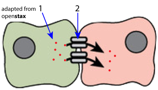
Adjacent animal cells can have gap junctions (at “2” in figure 7) that allow substances — including signaling molecules (1) —to move from the cytoplasm of one cell to the next.
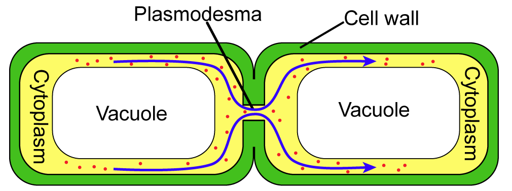
Adjacent plant cells have cytoplasmic connections called plasmodesmata. Like gap junctions, these allow signaling molecules (along with water and other substances) to freely diffuse from cell to cell.
Another form of cell communication by direct contact involves cells binding to one another through membrane receptors.
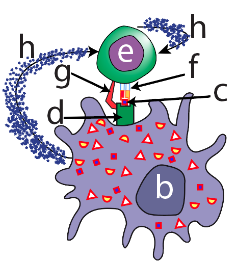
For example, the image at the left shows a key step in our immune response. A cell called a macrophage (at “b”) is communicating with a Helper T Cell (at “e”). The macrophage is a sentinel: it patrols our tissues, looking for infectious pathogens (disease-causing agents). When it finds one, it displays a piece of it (“c”) on a special protein (at “d”). In effect, the macrophage is saying “We’ve been invaded. Here’s what it looks like. Let’s counterattack.”
The counterattack only becomes possible when the macrophage meets a Helper T cell with a T cell receptor (“f”) that has a shape that’s complementary to a surface molecule from the invading pathogen that’s being displayed by the macrophage. This meeting will occur in a lymph node, and once it does, the macrophage will signal the Helper T cell with chemical messages called cytokines (at “H”).
If you look at the word cytokine, you’ll see the beginning of “cytokinesis.” Cytokinesis means “cell division.” Cytokines induce the Helper T cell to clone itself into millions of copies that will coordinate an assault on the invader.
The connection between a macrophage and a helper-T cell also shows us another aspect of cell communication. Cells can release ligands that induce changes not only in other cells but also in themselves. This kind of signaling is called autocrine signaling, and you can see it in the top right of Figure 9 above.
5b. Paracrine Signaling
In local signaling, cells are close to one another but aren’t directly touching.
One form of local signaling is called paracrine signaling. In paracrine signaling, one cell (or a group of cells) secretes signaling molecules that are received by receptors in nearby cells, changing those cells’ activities.
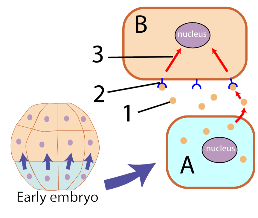
Paracrine signaling is a key process during animal development. In Figure 10 on the left, we see cells (“A”) from a very early embryo releasing a type of signaling molecule called a morphogen (“1”). These molecules are called morphogens because they control the development of form (morphology is the study of form). The morphogen is a ligand that binds with a receptor (“2”) in another cell (“B”). Signal transduction relays the message to the nucleus, activating genes that will determine how those cells will develop.
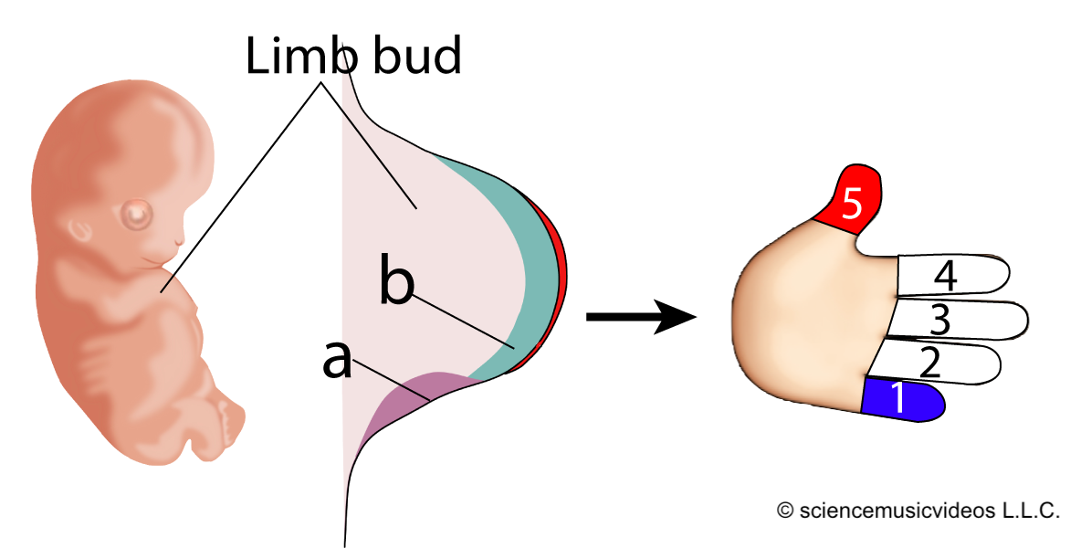
Here’s a more specific example. Before we develop a fully articulated arm and hand, we have a relatively undifferentiated limb bud. A bit of tissue in that limb bud (at “a”) secretes morphogens (“b”) that are received by other cells in the limb bud. Based on the concentration of the morphogen received, the cells are induced to develop into a pinky (“1”), a thumb (“5”), or the digits in between.
5c. Synaptic Signaling
A specialized form of local signaling occurs at a synapse, the connection between a neuron and its target.
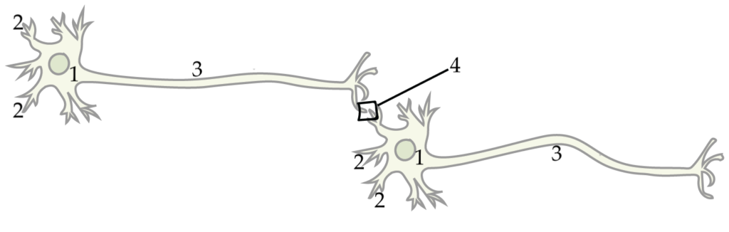
A nerve cell has a cell body (at “1”), and a long extension called an axon (“3”) through which a neuron sends its signal. At the end of the axon, the neuron doesn’t directly touch its target. Rather, there’s a gap called a synapse, shown above at “4.”
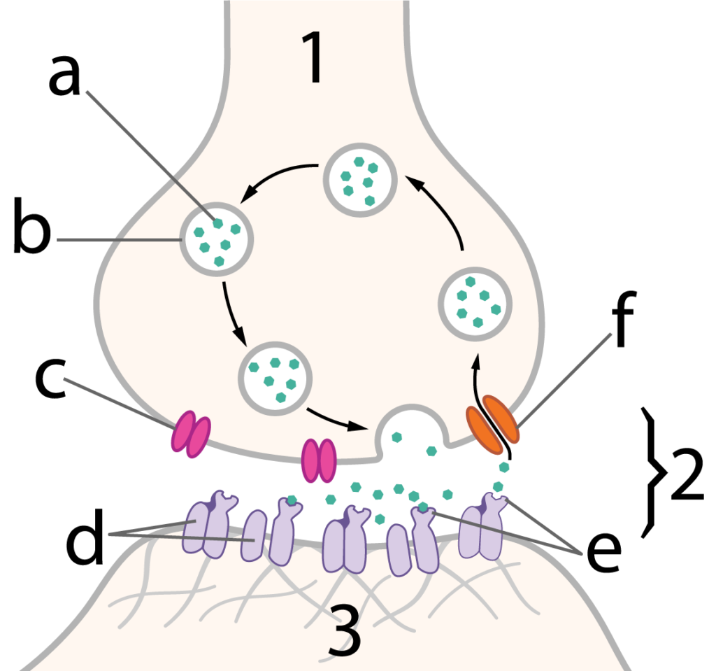
Figure 13 shows a synapse in detail. Number “1” shows the tip of an axon. Number “3” shows a postsynaptic cell, and “2” shows the synapse.
When a nerve impulse gets to the end of the axon, changes in membrane channels (“c”) induce vesicles (“b”) filled with neurotransmitter (“a”) to fuse with the tip of the axon. This releases neurotransmitters into the synapse.
All neurotransmitters are ligands. Once released, they diffuse across the synapse and bind with receptors (“e”) in a postsynaptic cell. Often, these receptors are connected to channels, which open upon binding, allowing ions to enter the postsynaptic cell. The effect depends on the neurotransmitter. Some are stimulatory and result in muscle contraction or another nerve impulse. Others are inhibitory.
5d. Long Distance (Endocrine) Signaling
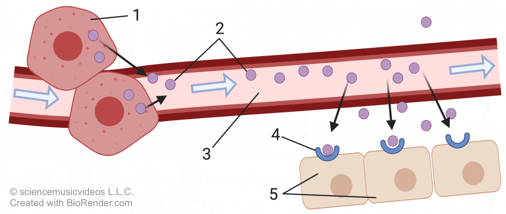
In multicellular organisms, long-distance signaling involves hormones (2), secreted by glands (“1”). These hormones diffuse into the bloodstream, where they travel throughout the entire body.
Cells (“5”) with receptors (“4”) specific to that hormone will respond by changing their activities.
While it’s hard to see this in Figure 14, the distance might be from your brain to your toes (which is why this is called “long-distance” signaling).
We’ll look at the specific mechanisms of endocrine signaling in the next tutorials in this module. For now, let’s consolidate what you’ve read above.
6. Cell Communication Overview: Checking Understanding
[qwiz random = “true” qrecord_id=”sciencemusicvideosMeister1961-Cell Communication Overview 2 (local, auto, para, endocrine signaling)”]
[h]Cell Communication Overview Quiz 2
[i]
[q]The diagram below illustrates how [hangman] [hangman] works. If the [hangman] molecule (at “2”) is at low density, then the [hangman] (at “3”) will remain unbound, and no cellular response will result. However, when there are a lot of signaling molecules, then they will bind with the receptor. In this case, the result is the activation of specific [hangman], changing cellular activities.
[c]cXVvcnVt[Qq]
[c]c2Vuc2luZw==[Qq]
[c]c2lnbmFs[Qq]
[c]cmVjZXB0b3I=[Qq]
[c]Z2VuZXM=[Qq]
[q]The structure at “3” is one of several [hangman] that connect plant cells (note: use the plural form)
[c]cGxhc21vZGVzbWF0YQ==[Qq]
[q]The structure at “2” is a [hangman] [hangman].
[c]Z2Fw[Qq]
[c]anVuY3Rpb24=[Qq]
[q]In the diagram below, a macrophage is displaying a molecule from a pathogen to a helper T cell. This type of cell communication is based on the shape of the T cell’s [hangman] (shown at “f”).
[c]cmVjZXB0b3I=[Qq]
[q]In the diagram below, the letter “h” appears twice. On the upper right side, “h” is an example of [hangman] signaling.
[c]YXV0b2NyaW5l[Qq]
[q] In the diagram below, which number or letter indicates a morphogen?
[textentry single_char=”true”]
[c]ID E=[Qq]
[f]IFdheSB0byBnbyEgTnVtYmVyICYjODIyMDsxJiM4MjIxOyByZXByZXNlbnRzIGEgbW9ycGhvZ2VuIChhIG1vbGVjdWxlIHRoYXQgaW5mbHVlbmNlcyBkZXZlbG9wbWVudCku[Qq]
[c]IEVudGVyIHdvcmQ=[Qq]
[c]ICo=[Qq]
[f]IE5vLiBIZXJlJiM4MjE3O3MgYSBoaW50LiBBIG1vcnBob2dlbiAoYSBtb2xlY3VsZSB0aGF0IGluZmx1ZW5jZXMgZGV2ZWxvcG1lbnQpIGlzIGEgc2lnbmFsaW5nIG1vbGVjdWxlLg==[Qq]
[q] In the diagram below, which number or letter indicates a receptor?
[textentry single_char=”true”]
[c]ID I=[Qq]
[f]wqAgRmFidWxvdXMhIE51bWJlciAmIzgyMjA7MiYjODIyMTsgcmVwcmVzZW50cyBhIHJlY2VwdG9yLg==[Qq]
[c]IEVudGVyIHdvcmQ=[Qq]
[c]ICo=[Qq]
[f]IE5vLiBIZXJlJiM4MjE3O3MgYSBoaW50LiBUaGUgcmVjZXB0b3IgaXMgd2hhdCB0aGUgc2lnbmFsaW5nIG1vbGVjdWxlIGJpbmRzIHdpdGgu[Qq]
[q]The diagram below is an example of [hangman] signaling
[c]cGFyYWNyaW5l[Qq]
[q]The diagram below is an example of [hangman] signaling.
[c]c3luYXB0aWM=[Qq]
[q] In the diagram below, which number or letter indicates a neurotransmitter?
[textentry single_char=”true”]
[c]IG M=[Qq]
[f]wqAgRmFidWxvdXMhIExldHRlciAmIzgyMjA7YyYjODIyMTsgcmVwcmVzZW50cyBhIG5ldXJvdHJhbnNtaXR0ZXIu[Qq]
[c]IEVudGVyIHdvcmQ=[Qq]
[c]ICo=[Qq]
[f]IE5vLiBIZXJlJiM4MjE3O3MgYSBoaW50LiBUaGUgbmV1cm90cmFuc21pdHRlciBpcyB0aGUgc2lnbmFsaW5nIG1vbGVjdWxlIHRoYXQgZGlmZnVzZXMgZnJvbSB0aGUgcHJlc3luYXB0aWMgbmV1cm9uIChhdCAmIzgyMjA7MSYjODIyMTspLCBhY3Jvc3MgdGhlIHN5bmFwc2UgKCYjODIyMDsyJiM4MjIxOyku[Qq]
[q] In the diagram below, which number or letter indicates the synapse?
[textentry single_char=”true”]
[c]ID I=[Qq]
[f]wqAgRXhjZWxsZW50ISBOdW1iZXIgJiM4MjIwOzImIzgyMjE7IHJlcHJlc2VudHMgdGhlIHN5bmFwc2Uu[Qq]
[c]IEVudGVyIHdvcmQ=[Qq]
[c]ICo=[Qq]
[f]IE5vLiBIZXJlJiM4MjE3O3MgYSBoaW50LiBUaGUgc3luYXBzZSBpcyB0aGUgZ2FwIGJldHdlZW4gdGhlIHByZXN5bmFwdGljIGF4b24gKGF0ICYjODIyMDsxJiM4MjIxOykgYW5kIHRoZSBwb3N0c3luYXB0aWMgZGVuZHJpdGUgKGF0ICYjODIyMDszLiYjODIyMTsp[Qq]
[q]The diagram below represents [hangman] signaling. In this system, signaling molecules called [hangman] (at “a”) are stored in [hangman] (at “b”) until a nerve impulse causes these signals to be released outside the cell. Once released, they diffuse across a [hangman] (at “2”) until they bind with [hangman] (at “e”), inducing changes in the postsynaptic cell (at “3”).
[c]c3luYXB0aWM=[Qq]
[c]bmV1cm90cmFuc21pdHRlcnM=[Qq]
[c]dmVzaWNsZXM=[Qq]
[c]c3luYXBzZQ==[Qq]
[c]cmVjZXB0b3Jz[Qq]
[q] In the diagram below, which number represents autocrine signaling?
[textentry single_char=”true”]
[c]ID E=[Qq]
[f]wqAgTmljZSEgTnVtYmVyICYjODIyMDsxJiM4MjIxOyByZXByZXNlbnRzIGF1dG9jcmluZSBzaWduYWxpbmcu[Qq]
[c]IEVudGVyIHdvcmQ=[Qq]
[c]ICo=[Qq]
[f]IE5vLiBIZXJlJiM4MjE3O3MgYSBoaW50LiBJbiBhdXRvY3JpbmUgc2lnbmFsaW5nLCBhIGNlbGwgc2VuZHMgb3V0IG1lc3NhZ2VzIHRoYXQgc3RpbXVsYXRlIGl0c2VsZi4=[Qq]
[q] In the diagram below, which number represents paracrine signaling?
[textentry single_char=”true”]
[c]ID M=[Qq]
[f]wqAgR29vZCB3b3JrISBOdW1iZXIgJiM4MjIwOzMmIzgyMjE7IHJlcHJlc2VudHMgcGFyYWNyaW5lIHNpZ25hbGluZy4=[Qq]
[c]IEVudGVyIHdvcmQ=[Qq]
[c]ICo=[Qq]
[f]IE5vLiBIZXJlJiM4MjE3O3MgYSBoaW50LiBJbiBwYXJhY3JpbmUgc2lnbmFsaW5nLCBhIGNlbGwgc2VuZHMgb3V0IG1lc3NhZ2VzIHRoYXQgZGlmZnVzZSB0byBhIG5lYXJieSBjZWxsLg==[Qq]
[q] In the diagram below, which number represents endocrine signaling?
[textentry single_char=”true”]
[c]ID Q=[Qq]
[f]IEF3ZXNvbWUhIE51bWJlciAmIzgyMjA7NCYjODIyMTsgcmVwcmVzZW50cyBlbmRvY3JpbmUgc2lnbmFsaW5nLg==[Qq]
[c]IEVudGVyIHdvcmQ=[Qq]
[c]ICo=[Qq]
[f]IE5vLiBIZXJlJiM4MjE3O3MgYSBoaW50LiBJbiBlbmRvY3JpbmUgc2lnbmFsaW5nLCBhIGNlbGwgc2VuZHMgb3V0IG1lc3NhZ2VzIHRoYXQgYXJlIHJlbGVhc2VkIGludG8gdGhlIGJsb29kc3RyZWFtIGFuZCB3aGljaCB0cmF2ZWwgdGhyb3VnaG91dCB0aGUgYm9keSw=[Qq]
[q]The diagram below shows four different types of signaling systems used by multicellular organisms. Diagram “1” shows [hangman] signaling. In this system, glands release [hangman] into the bloodstream. The only cells that are affected are those with the matching [hangman]. Diagram “2” shows a type of local signaling known as [hangman] signaling. In diagram 3, a cell is signaling itself, a kind of signaling called [hangman] signaling. In diagram 4, the cells are communicating by direct [hangman].
[c]ZW5kb2NyaW5l[Qq]
[c]aG9ybW9uZXM=[Qq]
[c]cmVjZXB0b3I=[Qq]
[c]cGFyYWNyaW5l[Qq]
[c]YXV0b2NyaW5l[Qq]
[c]Y29udGFjdA==[Qq]
[x][restart]
[/qwiz]
Links
- Continue to Cell Signaling (the next tutorial in this unit).
- Return to the Cell Communication Main Menu