Note from Mr. W. While the term “membrane-bound ribosome” is found in the College Board’s 2025 Course and Exam Description, most of the material in this tutorial is outside the scope of that document. Nevertheless, the biological principles are important (and highly recommended).
1. Introduction: Free vs. Bound Ribosomes

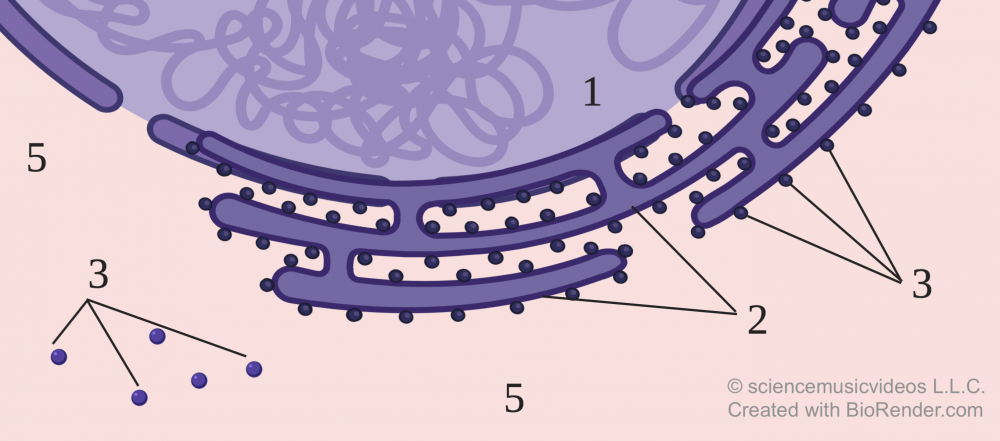
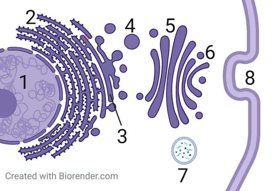
In a eukaryotic cell, ribosomes can be found in two places: in the cytoplasm (shown at “5” above), or bound to the rough endoplasmic reticulum (also known as the rough ER, shown at “2”). Ribosomes that are floating in the cytoplasm are called free ribosomes, and are indicated by “3.” The ribosomes attached to the rough ER are called bound ribosomes, and are shown at “4.”

When a free ribosome synthesizes a protein, it will release that protein directly into the cytoplasm. By contrast, bound ribosomes make proteins that are released into the ER lumen (the fluid space inside the ER). Afterward, these proteins will pass from the rough ER to the smooth ER (“3,” at right). This will be followed by transfers to vesicles (“4”), then the Golgi Apparatus (“5”), then more vesicles (“6”).
In the diagram above, several possible fates for these proteins are possible: the protein might integrate itself into the membrane, or the protein might be exported by the cell, or the protein might be encapsulated within a membrane-bound vesicle like a lysosome (shown at “7”).
Before going on, interact with these diagrams to make sure that you understand them. Doing so will help you review the endomembrane system, and also prepare you to understand protein targeting below.
[qwiz random=”true” qrecord_id=”sciencemusicvideosMeister1961-ER and Endomembrane System Review Quiz (2.0)”] [h]
The ER and Endomembrane System (Review)
[i]
[q] The nucleus is at
[textentry single_char=”true”]
[c]ID E=[Qq]
[f]IFllcy4gVGhlIG51Y2xldXMgaXMgYXQgJiM4MjIwOzEuJiM4MjIxOw==[Qq]
[c]IEVudGVyIHdvcmQ=[Qq]
[f]IE5vLg==[Qq]
[c]ICo=[Qq]
[f]IE5vLiBUaGUgbnVjbGV1cyBpcyB0aGUgbGFyZ2UsIGRhcmsgYm9keSBvbiB0aGUgZmFyIGxlZnQu[Qq]
[q] The rough ER is at
[textentry single_char=”true”]
[c]ID I=[Qq]
[f]IFllcy4gVGhlIHJvdWdoIEVSIGlzIGF0ICYjODIyMDsyLiYjODIyMTs=[Qq]
[c]IEVudGVyIHdvcmQ=[Qq]
[f]IE5vLg==[Qq]
[c]ICo=[Qq]
[f]IE5vLiBUaGUgcm91Z2ggRVIgaXMgYSBzZXJpZXMgb2YgbWVtYnJhbmUtYm91bmQgY2hhbm5lbHMgc3R1ZGRlZCB3aXRoIHJpYm9zb21lcy4gVGhlIHJpYm9zb21lcyBhcmUgcmVwcmVzZW50ZWQgYnkgdGhlIGRhcmsgYmxhY2sgZG90cy4=[Qq]
[q] A vesicle moving materials from the smooth ER to the Golgi is at
[textentry single_char=”true”]
[c]ID Q=[Qq]
[f]IFllcy4gTnVtYmVyICYjODIyMDs0JiM4MjIxOyByZXByZXNlbnRzIGEgdmVzaWNsZSB0aGF0JiM4MjE3O3MgbW92aW5nIG1hdGVyaWFscyBmcm9tIHRoZSBzbW9vdGggRVIgdG8gdGhlIEdvbGdpLg==[Qq]
[c]IEVudGVyIHdvcmQ=[Qq]
[f]IE5vLg==[Qq]
[c]ICo=[Qq]
[f]IE5vLiBIZXJlJiM4MjE3O3MgYSBoaW50LiBGaXJzdCBmaW5kIHRoZSBHb2xnaSAoYSBzZXJpZXMgb2YgbWVtYnJhbmUtYm91bmQsIGZsYXR0ZW5lZCBzYWNzKS4gVGhlbiBmaW5kIGEgdmVzaWNsZSB0aGF0JiM4MjE3O3MgbW92aW5nIHRvd2FyZHMgaXQgZnJvbSB0aGUgc21vb3RoIEVSLg==[Qq]
[q] The Golgi is at…
[textentry single_char=”true”]
[c]ID U=[Qq]
[f]IFllcy4gVGhlIEdvbGdpIGlzIGF0ICYjODIyMDs1LiYjODIyMTs=[Qq]
[c]IEVudGVyIHdvcmQ=[Qq]
[f]IE5vLg==[Qq]
[c]ICo=[Qq]
[f]IE5vLiBUaGUgR29sZ2kgaXMgYSBzZXJpZXMgb2YgZmxhdHRlbmVkIHNhY3Mu[Qq]
[q] An “outbound” vesicle heading toward the membrane or an organelle immediately after the protein it is carrying has been processed by the Golgi is at
[textentry single_char=”true”]
[c]ID Y=[Qq]
[f]IFllcy4gTnVtYmVyICYjODIyMDs2JiM4MjIxOyBpcyBhIHZlc2ljbGUgd2l0aCBhIHByb3RlaW4gdGhhdCBoYXMganVzdCBiZWVuIHByb2Nlc3NlZCBieSB0aGUgR29sZ2kgYXBwYXJhdHVzLg==[Qq]
[c]IEVudGVyIHdvcmQ=[Qq]
[f]IFNvcnJ5LCB0aGF0JiM4MjE3O3Mgbm90IGNvcnJlY3Qu[Qq]
[c]ICo=[Qq]
[f]IE5vLiBGaW5kIHRoZSB2ZXNpY2xlIHRoYXQmIzgyMTc7cyBjbG9zZXN0IHRvIHRoZSBHb2xnaSwgYW5kIHdoaWNoIGlzIG9uIHRoZSBHb2xnaSYjODIxNztzIG91dGVyIHNpZGUu[Qq]
[q] The nucleus is at
[textentry single_char=”true”]
[c]ID E=[Qq]
[f]IFllcy4gTnVtYmVyICYjODIyMDsxJiM4MjIxOyBpcyB0aGUgbnVjbGV1cw==[Qq]
[c]IEVudGVyIHdvcmQ=[Qq]
[f]IFNvcnJ5LCB0aGF0JiM4MjE3O3Mgbm90IGNvcnJlY3Qu[Qq]
[c]ICo=[Qq]
[f]IE5vLiBUaGUgbnVjbGV1cyBpcyBnZW5lcmFsbHkgaW4gdGhlIGNlbnRlciBvZiB0aGUgY2VsbCwgc28gZmluZCB0aGUgcGFydCBvZiB0aGlzIGRpYWdyYW0gdGhhdCBpcyBtb3N0IGNlbnRyYWwgKHlvdSBtaWdodCBoYXZlIHRvIGltYWdpbmUgdGhlIGVudGlyZSBkaWFncmFtIGFzIGEgY2lyY2xlKS4=[Qq]
[q] The rough ER is at
[textentry single_char=”true”]
[c]ID I=[Qq]
[f]IEV4Y2VsbGVudC4gTnVtYmVyICYjODIyMDsyJiM4MjIxOyBpcyB0aGUgcm91Z2ggRVI=[Qq]
[c]IEVudGVyIHdvcmQ=[Qq]
[f]IFNvcnJ5LCB0aGF0JiM4MjE3O3Mgbm90IGNvcnJlY3Qu[Qq]
[c]ICo=[Qq]
[f]IE5vLiBSb3VnaCBFUiBpcyBhIHNlcmllcyBvZiBpbnRlcmNvbm5lY3RlZCBtZW1icmFuZS1ib3VuZCBjaGFubmVscywgc3R1ZGRlZCB3aXRoIHJpYm9zb21lcy4gV2hpY2ggbnVtYmVyZWQgaXRlbSBhYm92ZSBjb3VsZCBmaXQgdGhhdCBkZXNjcmlwdGlvbj8=[Qq]
[q] Free ribosomes are indicated by which number?
[textentry single_char=”true”]
[c]ID M=[Qq]
[f]IE5pY2UuIE51bWJlciAmIzgyMjA7MyYjODIyMTsgaW5kaWNhdGVzIGZvdXIgJiM4MjIwO2ZyZWUmIzgyMjE7IHJpYm9zb21lcywgZmxvYXRpbmcgaW4gdGhlIGN5dG9wbGFzbS4=[Qq]
[c]IEVudGVyIHdvcmQ=[Qq]
[f]IFNvcnJ5LCB0aGF0JiM4MjE3O3Mgbm90IGNvcnJlY3Qu[Qq]
[c]ICo=[Qq]
[f]IE5vLiBGcmVlIHJpYm9zb21lcyBmbG9hdCBmcmVlbHkgaW4gdGhlIGN5dG9wbGFzbS4gSWYgdGhlIGN5dG9wbGFzbSBpcyBudW1iZXIgNSwgd2hpY2ggcmlib3NvbWVzIChyZXByZXNlbnRlZCBieSBibHVlIHNwaGVyZXMpIHdvdWxkIHF1YWxpZnkgYXMgYmVpbmcgJiM4MjIwO2ZyZWU/JiM4MjIxOw==[Qq]
[q] Bound ribosomes are indicated by which number?
[textentry single_char=”true”]
[c]ID Q=[Qq]
[f]IEdyZWF0IGpvYi4gTnVtYmVyICYjODIyMDs0JiM4MjIxOyBpbmRpY2F0ZXMgdGhyZWUgJiM4MjIwO2JvdW5kJiM4MjIxOyByaWJvc29tZXMsIGVhY2ggYXR0YWNoZWQgdG8gdGhlIG1lbWJyYW5lIG9mIHRoZSByb3VnaCBFUi4=[Qq]
[c]IEVudGVyIHdvcmQ=[Qq]
[f]IFNvcnJ5LCB0aGF0JiM4MjE3O3Mgbm90IGNvcnJlY3Qu[Qq]
[c]ICo=[Qq]
[f]IE5vLiBGcmVlIHJpYm9zb21lcyBmbG9hdCBmcmVlbHkgaW4gdGhlIGN5dG9wbGFzbS4gSWYgdGhlIGN5dG9wbGFzbSBpcyBudW1iZXIgNSwgd2hpY2ggcmlib3NvbWVzIChyZXByZXNlbnRlZCBieSBibHVlIHNwaGVyZXMpIHdvdWxkIHF1YWxpZnkgYXMgYmVpbmcgJiM4MjIwO2ZyZWU/JiM4MjIxOw==[Qq]
[/qwiz]
2. How do proteins get made at the right ribosome (“free” or “bound”)?
Now let’s step back for a moment and think about how protein synthesis works. A molecule of mRNA is synthesized in the nucleus and enters the cytoplasm. In the cytoplasm, a ribosome assembles itself around that mRNA and starts synthesizing whatever protein that the mRNA is coding for. As we’ve just seen, some of these proteins (those headed for export, the membrane, or organelles like lysosomes) have to be made by ribosomes at the rough E.R. How does the cell ensure that proteins that need to be made at the rough ER get to bound ribosomes, as opposed to free ones?
Based on what you know about biology, speculate about a mechanism. You can write these down on any piece of paper, or your student learning guide for this module.
3. Protein targeting works through a signal polypeptide that binds to a signal recognition particle
Almost everything in biology works by touch and feel. Why does adenine bind with thymine? Because of complementary shapes. That’s also why enzymes bind with substrates, or why the anticodon on a tRNA can bind with its corresponding codon on messenger RNA.
The process by which proteins destined for the ER wind up at the ER happens through a similar mechanism. Study the diagram before reading the text that follows, and see if you can explain to yourself what’s happening. Then read the text to confirm your hypothesis. Note that when you read, you can scroll the text while keeping the image stationary.

The first thing to realize is that all ribosomes start as free ribosomes. It’s the protein that the ribosome is synthesizing that determines whether or not the ribosome will dock at the ER membrane and become a “bound” ribosome. Here’s how it works:
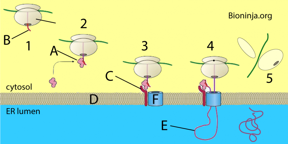
- STEP 1: At “1” you see a ribosome that’s starting to synthesize a protein. In ribosomes that are destined to make their proteins at the rough ER, the first few amino acids will make up a signal polypeptide, shown at “B.” The signal polypeptide will not be a part of the protein that’s released into the lumen of the rough ER (shown in step “5”). Rather, the signal polypeptide’s function is to bind with a signal recognition particle, shown by the letter “A.”
- STEP 2: At “2” you can see the signal recognition particle bound to the signal polypeptide. The signal recognition particle (SRP) has a second binding site (in addition to the one that binds with the signal polypeptide) that’s complementary to a translocation complex, a protein that’s embedded in the membrane of the rough ER. The translocation complex is indicated by the letters C and F. The binding of the SRP to the signal polypeptide also temporarily halts protein synthesis.
- STEP 3: In step 3, the ribosome is shown moving closer to the ER membrane (at “D.”) In diagrams like this the ribosome seems to know where to go, which of course is not the case. It’s important to keep in mind that the ribosome is mostly floating around randomly in the cytosol. When the ribosome, with its attached SRP, bumps into the ER’s membrane in the right spot and the right orientation, the SRP binds with the translocation complex (just as any ligand binds with its receptor). This is what you see in step “3.”
- STEP 4: At “4” you can see the signal polypeptide bound to the translocation complex. The binding of the signal polypeptide with the translocation complex restarts protein synthesis. with the rest of the polypeptide being threaded into the ER lumen. Note that the signal polypeptide will be enzymatically cleaved from the rest of the polypeptide, and not enter the ER lumen.
- STEP 5: At “5,” the ribosome has reached the stop codon on the mRNA. This causes the ribosomal subunits to dissociate, the mRNA to be released into the cytoplasm, and the protein to be released into the rough ER’s lumen. What’s not shown is that the SRP would be back in the cytoplasm, “waiting” to bind with another signal polypeptide. Enzymes in the cytoplasm will break the mRNA down into free nucleotides that can diffuse back into the nucleus where they can be reassembled back into various types of RNA.
Note that while our focus above has been on how ribosomes making certain proteins find their way to the rough ER, this kind of signaling is the basis for how many things find their right place within cells. Signal polypeptides are like the addresses on an envelope. These signals are bound by signal recognition particles that have affinities to specific organelles within the cell. In other words, protein targeting is how a cell sends proteins to its mitochondria, its lysosomes, or any other organelle.
Got it? Try this quiz.
4. Protein Targeting Quiz
[qwiz random = “true” style=”width: 600px !important;” qrecord_id=”sciencemusicvideosMeister1961-Protein Targeting Quiz (v2.0)”]
[h]Protein Targeting
[i]
[q] In the diagram below, a free ribosome that has synthesized a signal polypeptide, but has not yet bonded with a signal recognition particle, is shown at number
[textentry single_char=”true”]
[c]ID E=[Qq]
[f]IEV4Y2VsbGVudCEgJiM4MjIwOzEmIzgyMjE7IHNob3dzIGEgZnJlZSByaWJvc29tZS4gVGhpcyByaWJvc29tZSBoYXMgYWxyZWFkeSB0cmFuc2xhdGVkIHRoZSBzaWduYWwgcG9seXBlcHRpZGUsIGJ1dCB0aGF0IHNpZ25hbCBwb2x5cGVwdGlkZSBoYXMgbm90IHlldCBib25kZWQgd2l0aCB0aGUgc2lnbmFsIHJlY29nbml0aW9uIHBhcnRpY2xlLg==[Qq]
[c]IEVudGVyIHdvcmQ=[Qq]
[f]IFNvcnJ5LCB0aGF0JiM4MjE3O3Mgbm90IGNvcnJlY3Qu[Qq]
[c]ICo=[Qq]
[f]IE5vLiBIZXJlJiM4MjE3O3MgYSBoaW50LiBGcmVlIHJpYm9zb21lcyBhcmUgcmlib3NvbWVzIHRoYXQgaGF2ZW4mIzgyMTc7dCBkb2NrZWQgYXQgdGhlIEVSIG1lbWJyYW5lLiBPbmNlIHlvdSBmaW5kIGEgZnJlZSByaWJvc29tZSwgaG9uZSBpbiBvbiB0aGUgb25lIHRoYXQgaGFzbiYjODIxNzt0IGJvbmRlZCB3aXRoIHRoZSBzaWduYWwgcmVjb2duaXRpb24gcGFydGljbGUgKGF0ICYjODIyMDtBJiM4MjIxOyk=[Qq]
[q] In the diagram below, a free ribosome that has a signal recognition particle attached to its signal polypeptide is shown at number
[textentry single_char=”true”]
[c]ID I=[Qq]
[f]IE5pY2UhICYjODIyMDsyJiM4MjIxOyBzaG93cyBhIGZyZWUgcmlib3NvbWUuIFRoaXMgcmlib3NvbWUgaGFzIGFscmVhZHkgdHJhbnNsYXRlZCB0aGUgc2lnbmFsIHBvbHlwZXB0aWRlLCBhbmQgdGhlIHNpZ25hbCBwb2x5cGVwdGlkZSBoYXMgYm9uZGVkIHdpdGggdGhlIHNpZ25hbCByZWNvZ25pdGlvbiBwYXJ0aWNsZS4=[Qq]
[c]IEVudGVyIHdvcmQ=[Qq]
[f]IFNvcnJ5LCB0aGF0JiM4MjE3O3Mgbm90IGNvcnJlY3Qu[Qq]
[c]ICo=[Qq]
[f]IE5vLiBIZXJlJiM4MjE3O3MgYSBoaW50LiBGcmVlIHJpYm9zb21lcyBhcmUgcmlib3NvbWVzIHRoYXQgaGF2ZW4mIzgyMTc7dCBkb2NrZWQgYXQgdGhlIEVSIG1lbWJyYW5lLiBPbmNlIHlvdSBmaW5kIGEgZnJlZSByaWJvc29tZSwgaG9uZSBpbiBvbiB0aGUgb25lIHRoYXQmIzgyMTc7cyBib25kZWQgd2l0aCB0aGUgc2lnbmFsIHJlY29nbml0aW9uIHBhcnRpY2xlIChhdCAmIzgyMjA7QSYjODIyMTspLg==[Qq]
[q] In the diagram below, the first moment when a free ribosome becomes a bound ribosome is shown at number
[textentry single_char=”true”]
[c]ID M=[Qq]
[f]IEdvb2QgSm9iISAmIzgyMjA7MyYjODIyMTsgc2hvd3MgYSByaWJvc29tZSB0aGF0IGlzIGJvbmRpbmcgd2l0aCBhIHRyYW5zbG9jYXRpb24gY29tcGxleC4gVGhlIG1vbWVudCBhIGZyZWUgcmlib3NvbWUgYmluZHMgd2l0aCB0aGUgRVIgbWVtYnJhbmUsIGl0IGhhcyBiZWNvbWUgYSBib3VuZCByaWJvc29tZS4=[Qq]
[c]IEVudGVyIHdvcmQ=[Qq]
[f]IE5vLCB0aGF0JiM4MjE3O3Mgbm90IGNvcnJlY3Qu[Qq]
[c]ICo=[Qq]
[f]IE5vLiBIZXJlJiM4MjE3O3MgYSBoaW50LiBGcmVlIHJpYm9zb21lcyBhcmUgcmlib3NvbWVzIHRoYXQgaGF2ZW4mIzgyMTc7dCBkb2NrZWQgYXQgdGhlIEVSIG1lbWJyYW5lLiBJbiB0aGUgc2VxdWVuY2UgYWJvdmUsIGZpbmQgdGhlIG1vbWVudCB3aGVuIGEgZnJlZSByaWJvc29tZSBpcyBiaW5kaW5nIHdpdGggdGhlIEVSIG1lbWJyYW5lLg==[Qq]
[q] In the diagram below, the moment when the ribosome has dissociated, with ribosomal subunits and mRNA returning to the cytoplasm, is shown at number
[textentry single_char=”true”]
[c]ID U=[Qq]
[f]IENvcnJlY3QhICYjODIyMDs1JiM4MjIxOyBzaG93cyB0aGUgbW9tZW50IHdoZW4gdGhlIHJpYm9zb21lIGhhcyBkaXNzb2NpYXRlZC4=[Qq]
[c]IEVudGVyIHdvcmQ=[Qq]
[f]IE5vLCB0aGF0JiM4MjE3O3Mgbm90IGNvcnJlY3Qu[Qq]
[c]ICo=[Qq]
[f]IE5vLsKgIExvb2sgZm9yIHRoZSBtb21lbnQgd2hlbiB5b3Ugc2VlLCBpbiB0aGUgY3l0b3BsYXNtLCBib3RoIHRoZSBsYXJnZSBhbmQgc21hbGwgc3VidW5pdHMgb2YgdGhlIHJpYm9zb21lLCBhbG9uZyB3aXRoIGEgc3RyYW5kIG9mIG1STkEu[Qq]
[q] In the diagram below, “A” is a signal [hangman] particle.
[c]IHJlY29nbml0aW9u[Qq]
[f]IEdyZWF0IQ==[Qq]
[q] In the diagram below, “B” is a signal [hangman].
[c]IHBvbHlwZXB0aWRl[Qq]
[f]IEV4Y2VsbGVudCE=[Qq]
[q] In the diagram below, “C” and “F” make up the [hangman] complex.
[c]IHRyYW5zbG9jYXRpb24=[Qq]
[f]IEV4Y2VsbGVudCE=[Qq]
[q] In the diagram below, the signal recognition particle is shown at letter
[textentry single_char=”true”]
[c]IE E=[Qq]
[f]IENvcnJlY3QhICYjODIyMDtBJiM4MjIxOyBzaG93cyB0aGUgc2lnbmFsIHJlY29nbml0aW9uIHBhcnRpY2xl[Qq]
[c]IEVudGVyIHdvcmQ=[Qq]
[f]IE5vLCB0aGF0JiM4MjE3O3Mgbm90IGNvcnJlY3Qu[Qq]
[c]ICo=[Qq]
[f]IE5vLsKgIExvb2sgZm9yIGEgcGFydGljbGUgdGhhdCBiaW5kcyB3aXRoIHRoZSBzaWduYWwgcG9seXBlcHRpZGUgKHNob3duIGF0ICYjODIyMDtCLiYjODIyMTsp[Qq]
[q] In the diagram below, the signal polypeptide is shown at letter
[textentry single_char=”true”]
[c]IE I=[Qq]
[f]IFRlcnJpZmljISAmIzgyMjA7QiYjODIyMTsgc2hvd3MgdGhlIHNpZ25hbCBwb2x5cGVwdGlkZS4=[Qq]
[c]IEVudGVyIHdvcmQ=[Qq]
[f]IE5vLCB0aGF0JiM4MjE3O3Mgbm90IGNvcnJlY3Qu[Qq]
[c]ICo=[Qq]
[f]IE5vLsKgIExvb2sgZm9yIGEgcGllY2Ugb2YgdGhlIHBvbHlwZXB0aWRlIHRoYXQgdGhpcyByaWJvc29tZSBpcyBzeW50aGVzaXppbmcgdGhhdCAxKSBiaW5kcyB3aXRoIHRoZSBzaWduYWwgcmVjb2duaXRpb24gcGFydGljbGUsIGFuZCAyKcKgIGlzbiYjODIxNzt0IHVsdGltYXRlbHkgYSBwYXJ0IG9mIHRoZSBwcm90ZWluLg==[Qq]
[q] In the diagram below, what letter represents the protein in the ER membrane with which the signal recognition particle binds?
[textentry single_char=”true”]
[c]IE M=[Qq]
[f]IEF3ZXNvbWUhICYjODIyMDtDJiM4MjIxOyBzaG93cyB0aGUgcHJvdGVpbiBpbiB0aGUgdHJhbnNsb2NhdGlvbiBjb21wbGV4IHRoYXQgYWN0cyBhcyBhIGJpbmRpbmcgc2l0ZSBmb3IgdGhlIHNpZ25hbCByZWNvZ25pdGlvbiBwYXJ0aWNsZS4=[Qq]
[c]IEVudGVyIHdvcmQ=[Qq]
[f]IE5vLCB0aGF0JiM4MjE3O3Mgbm90IGNvcnJlY3Qu[Qq]
[c]ICo=[Qq]
[f]IE5vLsKgIExvb2sgZm9yIHdoYXQgbG9va3MgbGlrZSBhIHJlY2VwdG9yIHdpdGhpbiB0aGUgRVIgbWVtYnJhbmUu[Qq]
[q] In the diagram below, the rough ER Membrane is shown at letter
[textentry single_char=”true”]
[c]IE Q=[Qq]
[f]IEdvb2Qgd29yayEgJiM4MjIwO0QmIzgyMjE7IHNob3dzIHRoZSBFUiBtZW1icmFuZS4=[Qq]
[c]IEVudGVyIHdvcmQ=[Qq]
[f]IE5vLCB0aGF0JiM4MjE3O3Mgbm90IGNvcnJlY3Qu[Qq]
[c]ICo=[Qq]
[f]IE5vLsKgIExvb2sgZm9yIHNvbWV0aGluZyB0aGF0IGxvb2tzIGxpa2UgYSBtZW1icmFuZSBiZXR3ZWVuIHRoZSBjeXRvc29sIChpbiB3aGl0ZSkgYW5kIHRoZSBFUiBsdW1lbiAoaW4gYmx1ZSk=[Qq]
[q] In the diagram below, the protein that’s going to be released into the ER is shown at letter
[textentry single_char=”true”]
[c]IE U=[Qq]
[f]IEdvb2Qgam9iISAmIzgyMjA7RSYjODIyMTsgc2hvd3MgYSBwcm90ZWluIHRoYXQgaXMgZ29pbmcgdG8gYmUgcmVsZWFzZWQgaW50byB0aGUgRVIgbHVtZW4u[Qq]
[c]IEVudGVyIHdvcmQ=[Qq]
[f]IE5vLCB0aGF0JiM4MjE3O3Mgbm90IGNvcnJlY3Qu[Qq]
[c]ICo=[Qq]
[f]IE5vLsKgIExvb2sgZm9yIHNvbWV0aGluZyB0aGF0IGxvb2tzIGxpa2UgYSBtZW1icmFuZSBiZXR3ZWVuIHRoZSBjeXRvc29sIChpbiB3aGl0ZSkgYW5kIHRoZSBFUiBsdW1lbiAoaW4gYmx1ZSk=[Qq]
[q multiple_choice=”true”] Which of the following statements is true?
[c]IEFsbCByaWJvc29tZXMgc3RhcnQgZn JlZS4gU29tZSBiZWNvbWUgYm91bmQu[Qq]
[f]IFllcyEgQWxsIHJpYm9zb21lcyBiZWNvbWUgZnJlZS4gT25seSB0aG9zZSB0aGF0IHN5bnRoZXNpemUgYSBwcm90ZWluIHRoYXQgc3RhcnRzIHdpdGggYSBzaWduYWwgcG9seXBlcHRpZGUgYmVjb21lIGJvdW5kLg==[Qq]
[c]IEFsbCByaWJvc29tZXMgc3RhcnQgYXMgYm91bmQuIFNvbWUgYmVjb21lIGZyZWUu[Qq]
[f]IE5vLiBBbGwgcmlib3NvbWVzIHN0YXJ0IGZyZWUuIE9ubHkgdGhvc2UgdGhhdCBwcm9kdWNlIGEgcHJvdGVpbiB3aXRoIGEgc2lnbmFsIHJlY29nbml0aW9uIHBhcnRpY2xlIGJlY29tZSBib3VuZC4=[Qq]
[x][restart]
[/qwiz]
Next Moves
This tutorial ends AP Bio Topics 6.3 and 6.4, Transcription and Translation. Use the following link to access the next tutorial in AP Bio Unit 6, or use any of the menus above.
