Introduction
At this point, you should have a good understanding of homeostasis and adaptations for maintaining homeostasis through negative feedback. We’ll continue our study of homeostasis and feedback (AP Bio Topic 4.5) through an important illustrative example: blood glucose regulation. In the next tutorial, we’ll look at what happens when this system breaks down, causing diabetes.
1. Blood glucose levels fluctuate (but they’re still tightly controlled)
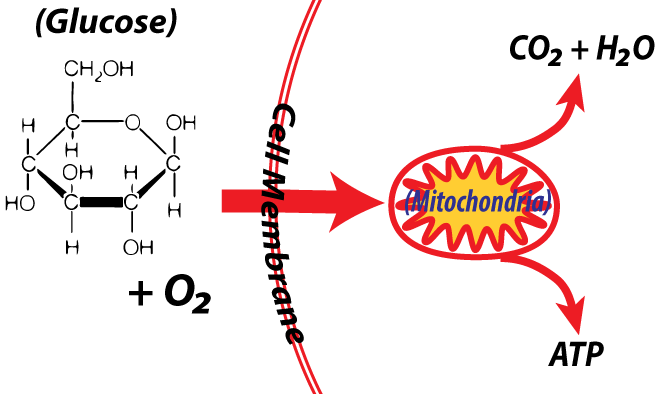
As you learned in AP Bio Unit 3, the monosaccharide glucose is essential to life because it’s a primary fuel that cells use to make ATP. For animals like us, the cells in our bodies acquire glucose from capillaries (tiny blood vessels) that supply the tissues of the body with blood. Glucose in the blood, in turn, comes from the food we eat, which gets absorbed into the blood through our intestines.
Unlike body temperature, which mammals keep confined to a narrow range, blood glucose levels can fluctuate by almost 50% throughout the day. Nevertheless, blood glucose levels are tightly regulated. Too little glucose in our glucose-hungry brains can send us into shock. Too much glucose is associated with diabetes, an increasingly common disease that affects about 30 million Americans (10% of the US population). Another 38% of the US population has a condition called prediabetes, a condition in which elevated blood glucose levels puts them at risk for diabetes later in life.
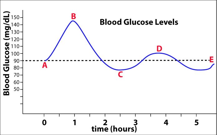 Blood glucose is measured in milligrams/deciliter (mg/dL). To put that in context, 100 mg/dL (which is close to the glucose set point) is just a few grains of sugar in a half a cup of water. Assuming that you’re not a diabetic or prediabetic, your set point for blood glucose is probably about 90 mg/dL (indicated by the dotted line at “E”).
After you eat (A), particularly if it’s a meal with starch or sugar, your blood glucose will rise to about 140 mg/dL (point “B”). At that point, homeostatic mechanisms will kick in, lowering your blood glucose back to its set point. If you go a few more hours without eating, then your glucose level will fall below the set point. At a certain point (C) other homeostatic mechanisms will kick in to raise your blood glucose back to the set point. Your blood glucose will continue to oscillate around the set point until you eat again.
2. Glucose Regulation: Setting the Context
 To understand how our bodies regulate blood glucose concentration, you need to know a few organs, tissues, and cell types.
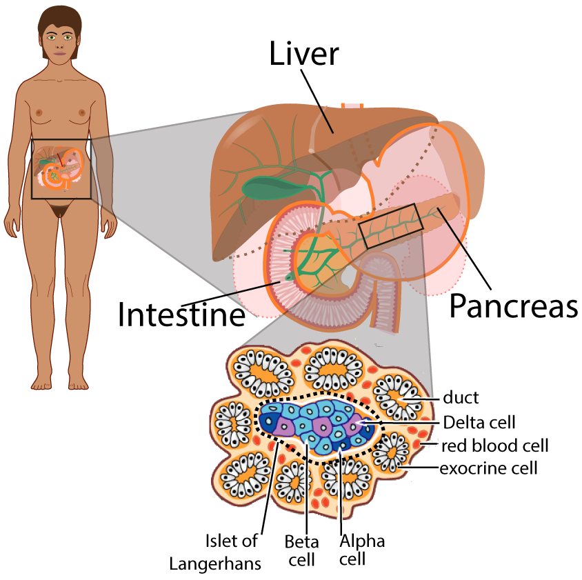
The pancreas (shown on the right) is just behind your stomach, high up in your abdomen. The pancreas mostly consists of tissues that create digestive enzymes and other digestive secretions. These are the exocrine cells in the callout on the right. These secretions are released into ducts that empty into the intestine.
In addition, the pancreas also consists of groups of cells called the Islets of Langerhans (named for their discoverer). These Islets contain two types of cells that release hormones that help control blood glucose levels.
Beta cells (colored light blue for clarity in the diagram) secrete the hormone insulin. The function of insulin is to lower blood glucose by inducing a variety of tissues to absorb glucose from the blood and convert it into the polysaccharide glycogen, or into fat. Alpha cells (colored dark blue) secrete glucagon, which induces various body tissues to break stored glycogen into glucose, which diffuses into the bloodstream, raising blood glucose levels. Delta cells produce a hormone that affects the stomach and are not involved in blood glucose regulation.
3. Insulin
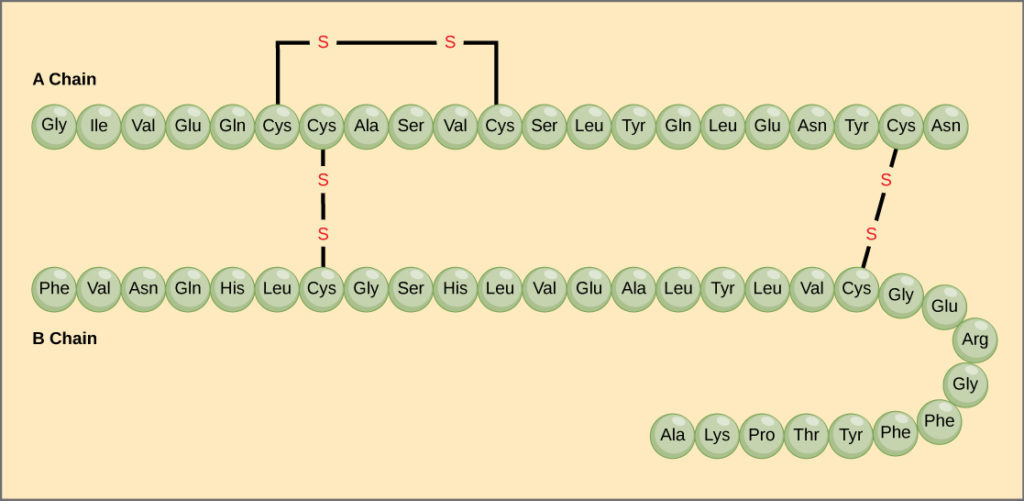
Insulin is a complex protein which usually has a quaternary structure. A single insulin unit (often referred to as an insulin monomer) consists of two polypeptide chains linked by disulfide bridges.
Secondary and tertiary interactions lead insulin to fold into a complex shape, and these molecules will interact with one another to form dimers (two units bonded together by hydrogen bonds) and hexamers (six units bonded together). The hexamer is used for storage, waiting to be released when the body needs to reduce blood glucose levels. The monomer is the most biologically active form.
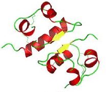
Insulin Dimer: Notice the alpha helices and the hairpin turns caused by side-chain interactions |
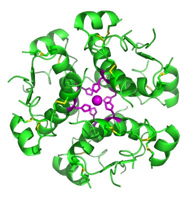
Insulin Hexamer: six bonded insulin units. |
4. Checking Understanding: Blood glucose, pancreatic hormones, and insulin
[qwiz random=”true” qrecord_id=”sciencemusicvideosMeister1961-Blood glucose, Pancreatic hormones, and Insulin (v2.0)”]
[h]Checking Understanding: Blood glucose, Pancreatic hormones, and Insulin
[i]
[q multiple_choice=”true”]In the diagram below, the effect of insulin can first be seen
[c]QmV0d2VlbiBBIGFuZCBC[Qq]
[f]Tm8uIEJldHdlZW4gQSBhbmQgQiBnbHVjb3NlIGxldmVscyBhcmUgcmlzaW5nLiBJbnN1bGluIGxvd2VycyBibG9vZCBnbHVjb3NlLg==[Qq]
[c]IEJldHdlZW4g QiBhbmQgQw==[Qq]
[f]IEV4Y2VsbGVudC4gVGhlIGxpbmUgYmV0d2VlbiAmIzgyMjA7QiBhbmQgQyYjODIyMTsgc2hvd3MgYmxvb2QgZ2x1Y29zZSBkcm9wcGluZyBhZnRlciBhIG1lYWwsIGFuIGVmZmVjdCB0aGF0IHdvdWxkIGJlIGJyb3VnaHQgYWJvdXQgYnkgaW5zdWxpbi4=[Qq]
[c]QmV0d2VlbiBDIGFuZCBE[Qq]
[f]IE5vLiBIZXJlJiM4MjE3O3MgYSBoaW50LiBJbnN1bGluIGxvd2VycyBibG9vZCBzdWdhciBsZXZlbHMuIFdoZXJlIGRvIHlvdSBzZWUgdGhhdCBoYXBwZW5pbmc/[Qq]
[q]In the diagram below, at what point would you predict that glucagon would be released?
[textentry single_char=”true”]
[c]IE M=[Qq]
[f]IE5pY2UuICYjODIyMDtDJiM4MjIxOyBzaG93cyB0aGUgbG93ZXN0IHBvaW50IG9mIGJsb29kIGdsdWNvc2UsIGZvbGxvd2VkIGJ5IGEgcmlzZS4gVGhhdCByaXNlIHdvdWxkIGJlIGJyb3VnaHQgYWJvdXQgYnkgZ2x1Y2Fnb24u[Qq]
[c]IEVudGVyIHdvcmQ=[Qq]
[c]ICo=[Qq]
[f]IE5vLiBIZXJlJiM4MjE3O3MgYSBoaW50LiBHbHVjYWdvbiBpbmNyZWFzZXMgYmxvb2QgZ2x1Y29zZSBsZXZlbHMuIFdoZXJlIGRvIHlvdSBzZWUgdGhhdCBzdGFydGluZyB0byBoYXBwZW4/[Qq]
[q]In the diagram below, at what point would glucose first start to be converted into glycogen and fat?
[textentry single_char=”true”]
[c]IE I=[Qq]
[f]IE5pY2UuICYjODIyMDtCJiM4MjIxOyBzaG93cyB0aGUgaGlnaGVzdCBibG9vZCBnbHVjb3NlIGxldmVsLCBmb2xsb3dlZCBieSBhIGRyb3AuIFRoYXQgZHJvcCBvY2N1cnMgYXMgaW5zdWxpbiBpbmR1Y2VzIHRoZSBsaXZlciAoYW5kIG90aGVyIHRpc3N1ZXMpIHRvIGFic29yYiBnbHVjb3NlIGZyb20gdGhlIGJsb29kIGFuZCBjb252ZXJ0IGl0IGludG8gZ2x5Y29nZW4u[Qq]
[c]IEVudGVyIHdvcmQ=[Qq]
[c]ICo=[Qq]
[f]IE5vLiBIZXJlJiM4MjE3O3MgYSBoaW50LiBBcyBnbHVjb3NlIGlzIGNvbnZlcnRlZCBpbnRvIGdseWNvZ2VuIGFuZCBmYXQsIGJsb29kIGdsdWNvc2UgbGV2ZWxzIHdvdWxkIHN0YXJ0IGRyb3BwaW5nLiBBdCB3aGF0IHBvaW50IGRvIHlvdSBzZWUgdGhhdCBzdGFydGluZyB0byBoYXBwZW4/[Qq]
[q]In the diagram below, the glucose set point is represented by
[textentry single_char=”true”]
[c]IE U=[Qq]
[f]IE5pY2UuICYjODIyMDtFJiM4MjIxOyByZXByZXNlbnRzIHRoZSBzZXQgcG9pbnQu[Qq]
[c]IEVudGVyIHdvcmQ=[Qq]
[c]ICo=[Qq]
[f]IE5vLiBIZXJlJiM4MjE3O3MgYSBoaW50LiBUaGUgc2V0IHBvaW50IGlzIHRoZSBsZXZlbCB0aGF0IHRoZSBib2R5IGlzICYjODIyMDt0cnlpbmcmIzgyMjE7IHRvIGFjaGlldmUgdGhyb3VnaCB2YXJpb3VzIGhvbWVvc3RhdGljIG1lY2hhbmlzbXMuIFdoYXQmIzgyMTc7cyB0aGUgdmFsdWUgdGhhdCBldmVyeXRoaW5nIGluIHRoZSBkaWFncmFtIHNlZW1zIHRvIGJlIG9zY2lsbGF0aW5nIGFyb3VuZD8=[Qq]
[q]The organ that’s responsible for releasing the hormones that maintain blood glucose homeostasis is the [hangman]. The hormone that lowers blood glucose back to the set point is [hangman]. The hormone that raises blood glucose back to the set point is [hangman].
[c]cGFuY3JlYXM=[Qq]
[c]aW5zdWxpbg==[Qq]
[c]Z2x1Y2Fnb24=[Qq]
[q]To lower blood glucose levels, glucose in the blood is converted by cells in the liver and muscles into the polysaccharide [hangman] and [hangman], which is a lipid.
[c]Z2x5Y29nZW4=[Qq]
[c]ZmF0[Qq]
[q]Protein review: In the diagram below the spirals represent [hangman] [hangman], which are at the [hangman] level of protein structure. The hairpin turns represent a [hangman]-level interactions, and interaction between multiple folded proteins is an example of [hangman]-level structure
[c]YWxwaGE=[Qq]
[c]aGVsaWNlcw==[Qq]
[c]c2Vjb25kYXJ5[Qq]
[c]dGVydGlhcnk=[Qq]
[c]cXVhdGVybmFyeQ==[Qq]
[q]Protein review: The diagram below indicates how insulin is composed of two [hangman] chains, linked by [hangman] [hangman].
[c]cG9seXBlcHRpZGU=[Qq]
[c]ZGlzdWxmaWRl[Qq]
[c]YnJpZGdlcw==[Qq]
[x][restart]
[/qwiz]
5. Control of Insulin Release
How do the beta cells in the pancreas “know” when to release insulin? The short answer is that the presence of glucose unleashes a signaling cascade that results in insulin release. The long answer involves a complex signaling pathway that you, an AP Bio student, should be able to handle. Let’s take a look.
Start by finding glucose in the diagram on the left, at about 11 o’clock.
After a carbohydrate-rich meal, glucose will diffuse into the blood from the small intestine. From the blood, glucose will diffuse to the beta cells in the pancreas.
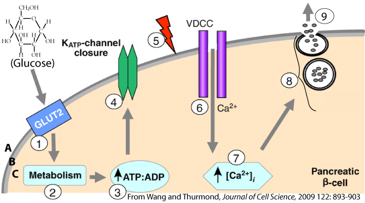
Because glucose is polar, it needs a channel to pass from the extracellular fluid (“A”) across the membrane (“B”) and into the cytoplasm (“C”). That channel is called GLUT2 (see “1”) and it allows glucose to enter the cytoplasm of the pancreatic Beta cells by facilitated diffusion.
Once in the cytoplasm, glucose becomes the substrate for cellular respiration (represented by “2”). Respiration increases cytoplasmic ATP (represented by the up arrow at “3”).
To understand what happens next you need to know that for a variety of reasons, cells have an electrical charge across their membrane. The outside of the cell has a positive charge, and the inside has a negative charge. This condition is called polarization.
What happens next causes this polarity to temporarily decrease. The ATP created by cellular respiration (at “3”) binds with a potassium channel (“4”) in the Beta cell’s membrane. Binding causes the channel to close. With the channel closed, positively charged potassium ions can no longer diffuse out of the cell. This reduces the charge across the membrane. That’s because these positively charged ions are now stuck inside the cell.
This reduced charge is called depolarization, and it’s represented by the red lightning bolt at “5.” A voltage-dependent calcium channel at “6” senses this change and responds by opening up. This allows calcium ions to diffuse into the cytoplasm where they interact with insulin-containing vesicles in such a way that induces exocytosis (“8”) releasing insulin into the bloodstream (“9”).
Note that parts of this mechanism are similar, in many respects, to what happens when nerve cells release neurotransmitters, or when muscle cells respond to nerve impulses by contracting.
6. Insulin’s Effect on Liver, Fat, and Muscle Tissue
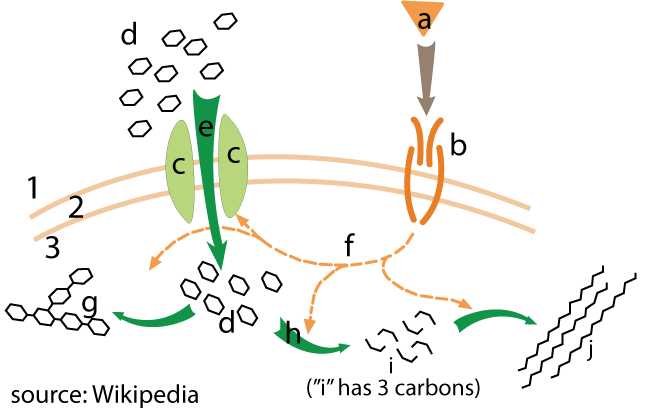 Once released from the pancreas, insulin will circulate throughout the body in the bloodstream. In the diagram on the right, insulin is represented by the letter “a.” Insulin will bind with receptors (“b”) on cells in your liver, muscles, and fatty tissues. Binding with the insulin receptor causes a signal transduction cascade (“f”) that has various effects on insulin’s target cells.
One of the most important of these effects is to bind with glucose transport channels (“c”). In response, these channels open, allowing for glucose (“d”) to enter the cells via facilitated diffusion (“e”).
Once inside the cytoplasm, enzymes might use dehydration synthesis to convert glucose into the polysaccharide glycogen (at “g”). Alternatively, glucose might undergo glycolysis (“h”), breaking it down into the 3-carbon molecule pyruvic acid (“i”). In fat cells, pyruvate might be converted into fatty acids (“j”), which can then be stored away as triglycerides.
7. Insulin: Checking Understanding
[qwiz random=”true” qrecord_id=”sciencemusicvideosMeister1961-Insulin (v2.0)”]
[h]Insulin: Checking Understanding
[i]
[q]In the diagram below, which number refers to processes like cellular respiration?
[textentry single_char=”true”]
[c]ID I=[Qq]
[f]IEV4Y2VsbGVudC4gJiM4MjIwOzImIzgyMjE7IGluY3JlYXNlcyB0aGUgcmVsYXRpdmUgYW1vdW50IG9mIEFUUCBpbiB0aGUgY2VsbCwgd2hpY2ggY2FuIG9ubHkgY29tZSBhYm91dCB0aHJvdWdoIGNlbGx1bGFyIHJlc3BpcmF0aW9uLg==[Qq]
[c]IEVudGVyIHdvcmQ=[Qq]
[c]ICo=[Qq]
[f]IE5vLiBIZXJlJiM4MjE3O3MgYSBoaW50LiBUaGluayBhYm91dCB3aGF0IGNlbGx1bGFyIHJlc3BpcmF0aW9uIGNyZWF0ZXMuIE5vdyBmaW5kIGEgcHJvY2VzcyBvbiB0aGUgZGlhZ3JhbSB0aGF0JiM4MjE3O3MgY3JlYXRpbmcgaXQu[Qq]
[q]In the diagram below, three channels are shown. Which one is connected with reducing the charge across the membrane (which can also be described as causing a partial depolarization)?
[textentry single_char=”true”]
[c]ID Q=[Qq]
[f]IE5pY2UuwqAgV2hlbiAmIzgyMjA7NCYjODIyMTsgY2xvc2VzLCBwb3Rhc3NpdW0gaW9ucyBjYW4gbm8gbG9uZ2VyIGRpZmZ1c2Ugb3V0IG9mIHRoZSBjZWxsLiBLZWVwaW5nIGEgcG9zaXRpdmUgY2hhcmdlIGxvY2tlZCBpbnNpZGUgdGhlIG1lbWJyYW5lIGRlcG9sYXJpemVzIHRoZSBtZW1icmFuZS4=[Qq]
[c]IEVudGVyIHdvcmQ=[Qq]
[c]ICo=[Qq]
[f]IE5vLiBIZXJlJiM4MjE3O3MgYSBoaW50LiBPbmUgb2YgdGhlIGNoYW5uZWxzIHNob3duIGlzIGtlZXBpbmcgdGhlIHBvc2l0aXZlIHBvdGFzc2l1bSBpb25zIGluc2lkZSB0aGUgbWVtYnJhbmUsIGNhdXNpbmcgdGhlIG1lbWJyYW5lIHRvIGRlcG9sYXJpemUuIExvb2sgYXQgd2hhdCBlYWNoIGNoYW5uZWwgZG9lcywgYW5kIHlvdSBzaG91bGQgc2VlIHRoZSBhbnN3ZXIu[Qq]
[q]In the diagram below, three channels are shown. Which one allows in the ion that induces exocytosis?
[textentry single_char=”true”]
[c]ID Y=[Qq]
[f]IE5pY2UuwqAgV2hlbiAmIzgyMjA7NiYjODIyMTsgb3BlbnMsIGNhbGNpdW0gaW9ucyBkaWZmdXNlIGludG8gdGhlIGNlbGwsIGluZHVjaW5nIGV4b2N5dG9zaXMgb2YgaW5zdWxpbiB2ZXNpY2xlcy4=[Qq]
[c]IEVudGVyIHdvcmQ=[Qq]
[c]ICo=[Qq]
[f]IE5vLiBIZXJlJiM4MjE3O3MgYSBoaW50LiBGaW5kIHRoZSBjaGFubmVsIHRoYXQmIzgyMTc7cyBhbGxvd2luZyBjYWxjaXVtIGlvbnMgaW4uIE9yLCBpZiB0aGF0IGRvZXNuJiM4MjE3O3QgaGVscCwganVzdCB3b3JrIGJhY2t3YXJkIGZyb20gJiM4MjIwOzguJiM4MjIxOw==[Qq]
[q]In the diagram below, insulin is being released at
[textentry single_char=”true”]
[c]ID k=[Qq]
[f]IEF3ZXNvbWUuwqAgTnVtYmVyICYjODIyMDs5JiM4MjIxOyBpcyBzaG93aW5nIHRoZSByZWxlYXNlIG9mIGluc3VsaW4u[Qq]
[c]IEVudGVyIHdvcmQ=[Qq]
[c]ICo=[Qq]
[f]IE5vLiBIZXJlJiM4MjE3O3MgYSBoaW50LiBJbnN1bGluIGlzIHJlbGVhc2VkIGJ5IHZlc2ljbGVzIHRocm91Z2ggZXhvY3l0b3Npcy4gV2hlcmUgaXMgdGhhdCBoYXBwZW5pbmc/[Qq]
[q]In numbers 8 and 9, a [hangman] is fusing with the membrane and releasing insulin from the cell. This is an example of a membrane process called [hangman].
[c]dmVzaWNsZQ==[Qq]
[c]ZXhvY3l0b3Npcw==[Qq]
[q]The channels at 1, 4, and 6 let polar or charged molecules move from higher to lower concentration, but only through a channel. That makes what’s happening at these channels examples of [hangman] [hangman].
[c]ZmFjaWxpdGF0ZWQ=[Qq]
[c]ZGlmZnVzaW9u[Qq]
[q]In the diagram below, insulin is at
[textentry single_char=”true”]
[c]IG E=[Qq]
[f]IEF3ZXNvbWUuwqAgTGV0dGVyICYjODIyMDthJiM4MjIxOyBpcyBpbnN1bGluLg==[Qq]
[c]IEVudGVyIHdvcmQ=[Qq]
[c]ICo=[Qq]
[f]IE5vLiBIZXJlJiM4MjE3O3MgYSBoaW50LiBJbnN1bGluIGlzIHRoZSBsaWdhbmQuIFdoZW4gaXQgYmluZHMgd2l0aCBpdHMgcmVjZXB0b3IsIGl0IGFsbG93cyBnbHVjb3NlIHRvIGRpZmZ1c2UgaW50byB0aGUgY2VsbC4=[Qq]
[q]In the diagram below, the insulin receptor is at
[textentry single_char=”true”]
[c]IG I=[Qq]
[f]IE5pY2UuwqAgTGV0dGVyICYjODIyMDtiJiM4MjIxOyBpcyB0aGUgaW5zdWxpbiByZWNlcHRvci4=[Qq]
[c]IEVudGVyIHdvcmQ=[Qq]
[c]ICo=[Qq]
[f]IE5vLiBIZXJlJiM4MjE3O3MgYSBoaW50LiBJbnN1bGluIGlzIHRoZSBsaWdhbmQuIFdoYXQgaXMgdGhpcyBsaWdhbmQgYmluZGluZyB3aXRoPw==[Qq]
[q]In the diagram below, facilitated diffusion of glucose is indicated by
[textentry single_char=”true”]
[c]IG U=[Qq]
[f]IE5pY2UuwqAgTGV0dGVyICYjODIyMDtlJiM4MjIxOyBzaG93cyBnbHVjb3NlIGVudGVyaW5nIGludG8gY2VsbHMgdGhyb3VnaCBhIHByb3RlaW4gY2hhbm5lbCwgd2hpY2ggaXMgd2hhdCBmYWNpbGl0YXRlZCBkaWZmdXNpb24gaXMgYWxsIGFib3V0Lg==[Qq]
[c]IEVudGVyIHdvcmQ=[Qq]
[c]ICo=[Qq]
[f]IE5vLiBIZXJlJiM4MjE3O3MgYSBoaW50LiBMb29rIGZvciBhIG1vbGVjdWxlIHRoYXQmIzgyMTc7cyBkaWZmdXNpbmcgaW50byB0aGUgY2VsbCB0aHJvdWdoIGEgcHJvdGVpbiBjaGFubmVsLiBUaGF0JiM4MjE3O3MgZmFjaWxpdGF0ZWQgZGlmZnVzaW9uLg==[Qq]
[q]In the diagram below, a signal transduction cascade is indicated by
[textentry single_char=”true”]
[c]IG Y=[Qq]
[f]IE5pY2UuwqAgTGV0dGVyICYjODIyMDtmJiM4MjIxOyBzaG93cyBhIHNlY29uZCBzaWduYWwgdGhhdCYjODIxNztzIGdvaW5nIHRvIHRoZSBjeXRvcGxhc20gYW5kIG90aGVyIHBhcnRzIG9mIHRoZSBtZW1icmFuZS4gVGhlc2Ugc2lnbmFscyBhcmUgdHJhbnNtaXR0ZWQgaW4gY2VsbHMgdGhyb3VnaCBzaWduYWwgdHJhbnNkdWN0aW9uIGNhc2NhZGVzLg==[Qq]
[c]IEVudGVyIHdvcmQ=[Qq]
[c]ICo=[Qq]
[f]IE5vLiBIZXJlJiM4MjE3O3MgYSBoaW50LiBXaGVuIGluc3VsaW4gKGEpIGJpbmRzIHdpdGggaXRzIHJlY2VwdG9yIChiKSwgdGhhdCYjODIxNztzIHRoZSBmaXJzdCBtZXNzYWdlLiBXaGF0IGFycm93IGluc2lkZSB0aGUgY3l0b3BsYXNtIHNob3dzIHRyYW5zbWlzc2lvbiBvZiBhIHNlY29uZCBzaWduYWw/[Qq]
[q]In the diagram below, glycogen formation is shown at
[textentry single_char=”true”]
[c]IG c=[Qq]
[f]IE5pY2UuwqAgTGV0dGVyICYjODIyMDtnJiM4MjIxOyBzaG93cyBnbHVjb3NlIG1vbGVjdWxlcyB0aGF0IGhhdmUgYmVlbiBjaGFpbmVkIHRvZ2V0aGVyIHRvIGZvcm0gZ2x5Y29nZW4u[Qq]
[c]IEVudGVyIHdvcmQ=[Qq]
[c]ICo=[Qq]
[f]IE5vLiBIZXJlJiM4MjE3O3MgYSBoaW50LiBMZXR0ZXIgJiM4MjIwO2QmIzgyMjE7IGlzIGdsdWNvc2UuIEdseWNvZ2VuIGlzIGEgcG9seW1lciBvZiBnbHVjb3NlLg==[Qq]
[q]In the diagram below, glycolysis is shown at
[textentry single_char=”true”]
[c]IG g=[Qq]
[f]IE5pY2UuwqAgTGV0dGVyICYjODIyMDtoJiM4MjIxOyBzaG93cyBnbHVjb3NlIG1vbGVjdWxlcyAod2hpY2ggaGF2ZSBzaXggY2FyYm9ucykgYmVpbmcgYnJva2VuIGRvd24gaW50byBhIDMtY2FyYm9uIG1vbGVjdWxlLiBUaGF0IG1vbGVjdWxlIHdvdWxkIGJlIHB5cnV2aWMgYWNpZCwgYW5kIGl0JiM4MjE3O3MgdGhlIHByb2R1Y3Qgb2YgZ2x5Y29seXNpcy4=[Qq]
[c]IEVudGVyIHdvcmQ=[Qq]
[c]ICo=[Qq]
[f]IE5vLiBIZXJlJiM4MjE3O3MgYSBoaW50LiBHbHljb2x5c2lzIHByb2R1Y2VzIHB5cnV2aWMgYWNpZCwgd2hpY2ggaXMgYSAzLWNhcmJvbiBtb2xlY3VsZS4gV2hhdCBwcm9jZXNzIGlzIHByb2R1Y2luZyBhIDMtY2FyYm9uIG1vbGVjdWxlPw==[Qq]
[/qwiz]
8. Glucagon
Glucagon is a peptide hormone. It consists of 29 amino acids that curl into an alpha helix, as shown below
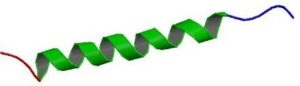
Glucagon works as insulin’s antagonist. Whereas insulin lowers blood glucose, glucagon does the opposite. It induces physiological responses in its target cells that raise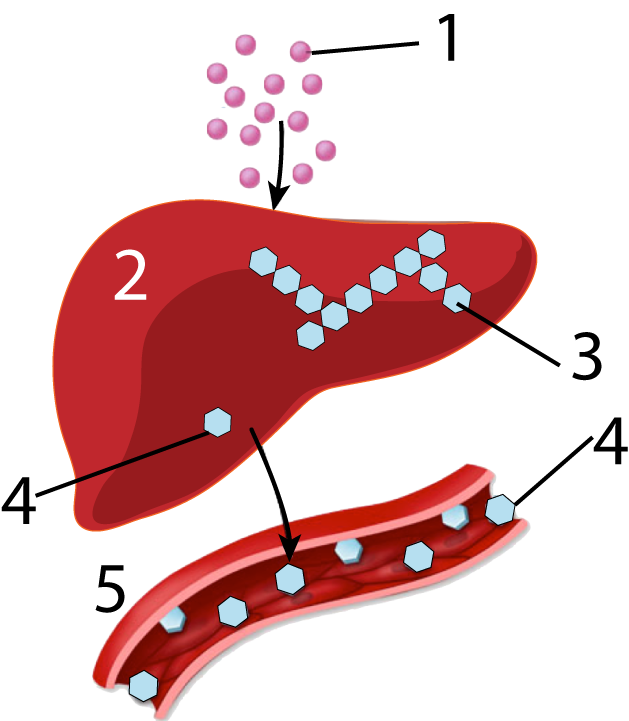 blood glucose levels toward the body’s set point.
You can see this happening in a general way in the diagram at right. Glucagon (1) is taken up by cells in the liver (2). In liver cells, a phosphorylation cascade activates enzymes (not shown) to hydrolyze the polysaccharide glycogen (3), which is a glucose polymer, into glucose. Next, glucose (4) can diffuse from the liver into the surrounding blood vessels (5), which increases the blood glucose level.
Earlier in this unit, you completed a series of tutorials about how epinephrine (also known as adrenaline) can mobilize tissue in the liver to break down the polysaccharide glycogen into glucose. Glucagon works through mechanisms that are similar, and, consequently, everything that follows is going to feel like a review.
Like epinephrine, glucagon binds with a G-protein-linked membrane receptor. Binding unleashes a chain of events at the membrane that result in the release of the second messenger, cyclic AMP (cAMP). Cyclic AMP, in turn, unleashes a phosphorylation cascade that results in the activation of the enzyme glycogen phosphorylase, which catalyzes the breakdown of glycogen into glucose.
Here are the details.
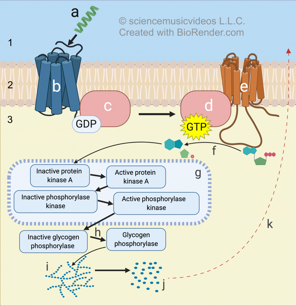
Glucagon (“a”) binds with a G-protein coupled receptor (“b”). Binding induces a conformational change in the G protein (“c”), which binds with GTP and releases GDP.
GTP activates the G protein, which now diffuses through the phospholipid bilayer (“2”) until it encounters the membrane-bound enzyme adenylyl cyclase (“e”). Adenylyl cyclase converts ATP into the second messenger, cyclic AMP (also known as cAMP). This conversion is represented by the arrow at “f.”
Cyclic AMP unleashes a phosphorylation cascade (“g”) in which protein kinases activate other kinases by adding phosphate groups to them. At the end of the chain, the target enzyme, glycogen phosphorylase a, is activated, and then converts glycogen (at “i”) to glucose (“j”), which can then diffuse out of the cell (indicated by the arrow at “k.”)
9. Summary: Two negative feedback loops maintain blood glucose homeostasis
Let’s step back from the cellular details and look at the entire system for maintaining blood glucose homeostasis.
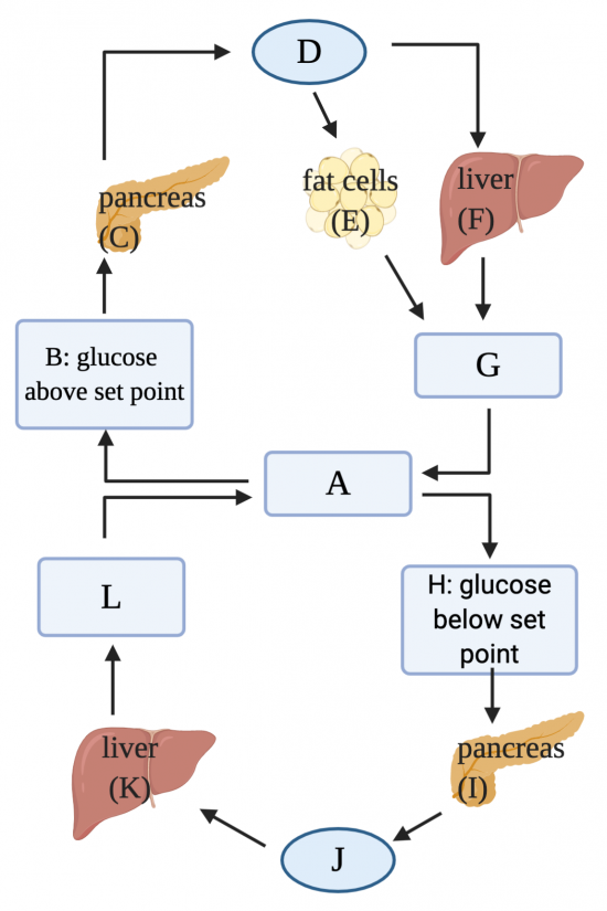
The glucose set point (about 90 mg/dL) is represented by “A.” After a meal, glucose will be absorbed from the intestines into the blood, causing the glucose level to rise above the set point (B). When this rise is detected by the Beta cells in the pancreas, they’ll release insulin (D), which induces fat cells (E) and cells in the liver (F) to take up glucose, lowering the blood glucose level (G) back to the set point.
A few hours after a meal, blood glucose levels will start to drop (H). When this drop is detected by alpha cells in the pancreas (I) glucagon is released (J). Glucagon acts on liver cells (K) and also muscle cells (not shown) to release glucose into the bloodstream, raising the blood glucose level (L) back toward the set point.
10. Insulin, Glucagon, and Blood Glucose Homeostasis: Checking Understanding
[qwiz qrecord_id=”sciencemusicvideosMeister1961-Insulin, Glucagon, and Blood Glucose Homeostasis (v2.0)”] [h]
Insulin, Glucagon, and Blood Glucose Homeostasis
[i]Biohaiku
Blood Glucose Levels
Insulin and Glucagon
Homeostasis
[q labels = “top”]
[l]Alpha cells release glucagon
[f*] Great!
[fx] No. Please try again.
[l]Beta cells release insulin
[f*] Excellent!
[fx] No. Please try again.
[l]Blood glucose falls
[f*] Good!
[fx] No. Please try again.
[l]Blood glucose rises
[f*] Great!
[fx] No. Please try again.
[l]Body cells take up glucose
[f*] Correct!
[fx] No, that’s not correct. Please try again.
[l]Liver breaks down glycogen, releases glucose
[f*] Correct!
[fx] No, that’s not correct. Please try again.
[l]Liver takes up glucose; makes glycogen
[f*] Good!
[fx] No, that’s not correct. Please try again.
[l]insulin
[f*] Great!
[fx] No, that’s not correct. Please try again.
[l]glucagon
[f*] Great!
[fx] No, that’s not correct. Please try again.
[q] In the diagram below, which letter shows where Beta cells would be releasing insulin?
[textentry single_char=”true”]
[c]IE M=[Qq]
[f]IEV4Y2VsbGVudC4gJiM4MjIwO0MmIzgyMjE7IHNob3dzIHRoZSBwYW5jcmVhcyByZWxlYXNpbmcgaW5zdWxpbiBpbiByZXNwb25zZSB0byBhIHJpc2UgaW4gYmxvb2QgZ2x1Y29zZS4=[Qq]
[c]IEVudGVyIHdvcmQ=[Qq]
[c]ICo=[Qq]
[f]IE5vLiBIZXJlJiM4MjE3O3MgYSBoaW50OiBpbnN1bGluIGlzIHJlbGVhc2VkIGJ5IGNlbGxzIGluIHRoZSBwYW5jcmVhcyBpbiByZXNwb25zZSB0byBhIHJpc2UgaW4gYmxvb2QgZ2x1Y29zZSBsZXZlbHMu[Qq]
[q] In the diagram below, where would glucose be converted into glycogen?
[textentry single_char=”true”]
[c]IE Y=[Qq]
[f]IEV4Y2VsbGVudC4gJiM4MjIwO0YmIzgyMjE7IGlzIHdoZXJlIHRoZSBsaXZlciB3b3VsZCBjb252ZXJ0IGdsdWNvc2UgdG8gZ2x5Y29nZW4u[Qq]
[c]IEVudGVyIHdvcmQ=[Qq]
[c]ICo=[Qq]
[f]IE5vLiBIZXJlJiM4MjE3O3MgYSBoaW50OiB0aGlzIGlzIG9uZSBvZiB0aGUgdHdvIG9yZ2FucyBzaG93biBhYm92ZSB0aGF0IGFyZSByZXNwb25kaW5nIHRvIGluc3VsaW4sIGFuZCB0aGlzIG9jY3VycyB3aGVuIGJsb29kIHN1Z2FyIGxldmVscyBhcmUgcmlzaW5nLg==[Qq]
[q] In the diagram below, what letter indicates insulin?
[textentry single_char=”true”]
[c]RA ==[Qq]
[f]IE5pY2Ugam9iLiAmIzgyMjA7RCYjODIyMTsgaGFzIHRvIGJlIGluc3VsaW46IHRoZSBob3Jtb25lIHRoYXQmIzgyMTc7cyByZWxlYXNlZCBpbiByZXNwb25zZSB0byByaXNpbmcgYmxvb2Qgc3VnYXIgbGV2ZWxzLg==[Qq]
[c]IEVudGVyIHdvcmQ=[Qq]
[c]ICo=[Qq]
[f]IE5vLiBIZXJlJiM4MjE3O3MgYSBoaW50OiBmaW5kIHdoZXJlIGJsb29kIGdsdWNvc2UgaXMgcmlzaW5nIGFib3ZlIHRoZSBzZXQgcG9pbnQuIEluc3VsaW4gaXMgdGhlIGhvcm1vbmUgcmVsZWFzZWQgaW4gcmVzcG9uc2Uu[Qq]
[q] In the diagram below, what letter indicates falling blood glucose levels?
[textentry single_char=”true”]
[c]Rw ==[Qq]
[f]IEF3ZXNvbWUuICYjODIyMDtHJiM4MjIxOyBpbmRpY2F0ZXMgdGhlIGJvZHkmIzgyMTc7cyByZXNwb25zZSB0byBpbnN1bGluIHJlbGVhc2U6IGZhbGxpbmcgYmxvb2QgZ2x1Y29zZSBsZXZlbHMu[Qq]
[c]IEVudGVyIHdvcmQ=[Qq]
[c]ICo=[Qq]
[f]IE5vLiBIZXJlJiM4MjE3O3MgYSBoaW50OiBibG9vZCBnbHVjb3NlIGxldmVscyB3aWxsIGZhbGwgaW4gcmVzcG9uc2UgdG8gdGhlIHJlbGVhc2Ugb2YgaW5zdWxpbiwgd2hpY2ggaXMgcmVsZWFzZWQgaW4gcmVzcG9uc2UgdG8gcmlzaW5nIGJsb29kIGdsdWNvc2UgbGV2ZWxzLg==[Qq]
[q] In the diagram below, what letter indicates glucagon?
[textentry single_char=”true”]
[c]Sg ==[Qq]
[f]IEF3ZXNvbWUuICYjODIyMDtKJiM4MjIxOyBpbmRpY2F0ZXMgZ2x1Y2Fnb24=[Qq]
[c]IEVudGVyIHdvcmQ=[Qq]
[c]ICo=[Qq]
[f]IE5vLiBIZXJlJiM4MjE3O3MgYSBoaW50OiBnbHVjYWdvbiBpcyByZWxlYXNlZCBpbiByZXNwb25zZSB0byByaXNpbmcgYmxvb2QgZ2x1Y29zZSBsZXZlbHMu[Qq]
[q] In the diagram below, what letter indicates where glycogen is being broken down to glucose?
[textentry single_char=”true”]
[c]Sw ==[Qq]
[f]IE5pY2UuICYjODIyMDtLJiM4MjIxOyBpbmRpY2F0ZXMgd2hlcmUgdGhlIGxpdmVyIHdvdWxkIGJlIGJyZWFraW5nIGRvd24gZ2x5Y29nZW4gdG8gZ2x1Y29zZS4=[Qq]
[c]IEVudGVyIHdvcmQ=[Qq]
[c]ICo=[Qq]
[f]IE5vLiBIZXJlJiM4MjE3O3MgYSBoaW50OiB0aGlzIGlzIGdvaW5nIHRvIG9jY3VyIGluIHJlc3BvbnNlIHRvIHRoZSByZWxlYXNlIG9mIGdsdWNhZ29uLCB3aGljaCBpcyBnb2luZyB0byBiZSByZWxlYXNlZCBieSB0aGUgcGFuY3JlYXMgaW4gcmVzcG9uc2UgdG8gZmFsbGluZyBibG9vZCBnbHVjb3NlIGxldmVscy4gU28sIHRoZSBxdWVzdGlvbiAoYW5kIGFuc3dlcikgaXMgd2hlcmU=IGlzIGl0IGdvaW5nIHRvIG9jY3VyPw==[Qq]
[q] In the diagram below, what letter indicates where Beta cells are releasing insulin?
[textentry single_char=”true”]
[c]Qw ==[Qq]
[f]IE5pY2Ugd29yay4gJiM4MjIwO0MmIzgyMjE7IGluZGljYXRlcyB3aGVyZSBiZXRhIGNlbGxzIGluIHRoZSBwYW5jcmVhcyB3b3VsZCBiZSByZWxlYXNpbmcgaW5zdWxpbi4=[Qq]
[c]IEVudGVyIHdvcmQ=[Qq]
[c]ICo=[Qq]
[f]IE5vLiBIZXJlJiM4MjE3O3MgYSBoaW50OiBCZXRhIGNlbGxzIHJlbGVhc2UgaW5zdWxpbi4gVGhpbmsgYWJvdXQgd2hhdCBvcmdhbiByZWxlYXNlcyBpbnN1bGluLCBhbmQgdW5kZXIgd2hhdCBjb25kaXRpb25zIGluc3VsaW4gaXMgcmVsZWFzZWQu[Qq]
[q] In the diagram below, what letter indicates where alpha cells are releasing glucagon?
[textentry single_char=”true”]
[c]SQ ==[Qq]
[f]IFdheSB0byBnby4gJiM4MjIwO0kmIzgyMjE7IGluZGljYXRlcyB3aGVyZSBhbHBoYSBjZWxscyBpbiB0aGUgcGFuY3JlYXMgd291bGQgYmUgcmVsZWFzaW5nIGdsdWNhZ29uLg==[Qq]
[c]IEVudGVyIHdvcmQ=[Qq]
[c]ICo=[Qq]
[f]IE5vLiBIZXJlJiM4MjE3O3MgYSBoaW50OiBBbHBoYSBjZWxscyBpbiB0aGUgcGFuY3JlYXMgcmVsZWFzZSBnbHVjYWdvbiwgd2hpY2ggaXMgcmVsZWFzZWQgd2hlbiBibG9vZCBnbHVjb3NlIGZhbGxzIGJlbG93IHRoZSBzZXQgcG9pbnQuIFB1dCB0aG9zZSB0d28gdG9nZXRoZXIsIGFuZCB5b3UmIzgyMTc7bGwgaGF2ZSB0aGUgYW5zd2VyLg==[Qq]
[q] In the diagram below, what letter indicates a G-protein coupled receptor?
[textentry single_char=”true”]
[c]Yg ==[Qq]
[f]IFdheSB0byBnby4gTGV0dGVyICYjODIyMDtiJiM4MjIxOyBpbmRpY2F0ZXMgdGhlIEctcHJvdGVpbiBjb3VwbGVkIHJlY2VwdG9yLg==[Qq]
[c]IEVudGVyIHdvcmQ=[Qq]
[c]ICo=[Qq]
[f]IE5vLiBIZXJlJiM4MjE3O3MgYSBoaW50OiBGaW5kIHRoZSBtZW1icmFuZS1lbWJlZGRlZCByZWNlcHRvciB0aGF0IGNhbiBiaW5kIHdpdGggYSBsaWdhbmQsIHdoaWNoIGluIHRoaXMgY2FzZSBpcyBnbHVjYWdvbi4=[Qq]
[q] In the diagram below, what letter indicates the inactive form of the G-protein?
[textentry single_char=”true”]
[c]Yw ==[Qq]
[f]IFdheSB0byBnby4gTGV0dGVyICYjODIyMDtjJiM4MjIxOyBpbmRpY2F0ZXMgdGhlIGluYWN0aXZlIGZvcm0gb2YgdGhlIEctcHJvdGVpbi4=[Qq]
[c]IEVudGVyIHdvcmQ=[Qq]
[c]ICo=[Qq]
[f]IE5vLiBIZXJlJiM4MjE3O3MgYSBoaW50OiBUaGUgRy1wcm90ZWluLCB3aGVuIGluYWN0aXZlLMKgIGlzIGxpbmtlZCB0byB0aGUgcmVjZXB0b3IsIHdoaWNoIGlzIGVtYmVkZGVkIGluIHRoZSBtZW1icmFuZS4=[Qq]
[q] In the diagram below, what letter indicates adenylyl cyclase?
[textentry single_char=”true”]
[c]ZQ ==[Qq]
[f]IE5pY2UuIExldHRlciAmIzgyMjA7ZSYjODIyMTsgaW5kaWNhdGVzIGFkZW55bHlsIGN5Y2xhc2Uu[Qq]
[c]IEVudGVyIHdvcmQ=[Qq]
[c]ICo=[Qq]
[f]IE5vLiBIZXJlJiM4MjE3O3MgYSBoaW50OiBBZGVueWx5bCBjeWNsYXNlIHJlc3BvbmRzIHRvIGEgc2lnbmFsIGZyb20gdGhlIEcgcHJvdGVpbiwgYW5kIGNvbnZlcnRzIEFUUCB0byBjeWNsaWMgQU1QLg==[Qq]
[q] In the diagram below, what letter shows the production of the second messenger?
[textentry single_char=”true”]
[c]Zg ==[Qq]
[f]IE5pY2UuIExldHRlciAmIzgyMjA7ZiYjODIyMTsgc2hvd3MgYWRlbnlseWwgY3ljbGFzZSBtYWtpbmcgQVRQIGludG8gY0FNUCwgd2hpY2ggaXMgdGhlIHNlY29uZCBtZXNzZW5nZXIu[Qq]
[c]IEVudGVyIHdvcmQ=[Qq]
[c]ICo=[Qq]
[f]IE5vLiBIZXJlJiM4MjE3O3MgYSBoaW50OiBUaGUgZW56eW1lIHRoYXQmIzgyMTc7cyBwcm9kdWNpbmcgdGhlIHNlY29uZCBtZXNzZW5nZXIgaXMgQWRlbnlseWwgY3ljbGFzZSwgYW5kIGl0JiM4MjE3O3MgY29udmVydGluZyBBVFAgaW50byB0aGUgc2Vjb25kIG1lc3NlbmdlciwgY0FNUC4=[Qq]
[q] In the diagram below, what letter shows a phosphorylation cascade?
[textentry single_char=”true”]
[c]Zw ==[Qq]
[f]IE5pY2UuIExldHRlciAmIzgyMjA7ZyYjODIyMTsgc2hvd3MgYSBwaG9zcGhvcnlsYXRpb24gY2FzY2FkZS4=[Qq]
[c]IEVudGVyIHdvcmQ=[Qq]
[c]ICo=[Qq]
[f]IE5vLiBIZXJlJiM4MjE3O3MgYSBoaW50LiBEdXJpbmcgYSBwaG9zcGhvcnlsYXRpb24gY2FzY2FkZSwgYSBzZXJpZXMgb2YgZW56eW1lcyBjYWxsZWQga2luYXNlcyBhY3RpdmF0ZSBlbnp5bWVzIGJ5IGFkZGluZyBwaG9zcGhhdGUgZ3JvdXBzIHRvIHRoZW0uIFdoZXJlIGluIHRoZSBkaWFncmFtIGRvIHlvdSBzZWUgdGhlIHdvcmQgJiM4MjIwO2tpbmFzZT8mIzgyMjE7[Qq]
[q] In the diagram below, where do you see the activation of the enzyme that converts glycogen into glucose?
[textentry single_char=”true”]
[c]aA ==[Qq]
[f]IEV4Y2VsbGVudC4gTGV0dGVyICYjODIyMDtoJiM4MjIxOyBzaG93cyB0aGUgY29udmVyc2lvbiBvZiBnbHljb2dlbiBwaG9zcGhvcnlsYXNlIGEgaW50byBwaG9zcGhvcnlsYXNlIGIsIGFuZCBwaG9zcGhvcnlsYXNlIGIgaXMgdGhlIGVuenltZSB0aGF0IGNvbnZlcnRzIGdseWNvZ2VuIGludG8gZ2x1Y29zZS4=[Qq]
[c]IEVudGVyIHdvcmQ=[Qq]
[c]ICo=[Qq]
[f]IE5vLiBIZXJlJiM4MjE3O3MgYSBoaW50LiBBdCB0aGUgYm90dG9tIG9mIHRoZSBkaWFncmFtLCB5b3Ugc2VlIGdseWNvZ2VuIGJlaW5nIGNvbnZlcnRlZCBpbnRvIGdsdWNvc2UuIFdoYXQgZW56eW1lIGlzIGNhdGFseXppbmcgdGhhdCBjb252ZXJzaW9uPw==[Qq]
[x][restart]
[/qwiz]
11. Blood Glucose Homeostasis Flashcards
[qdeck style=”width: 550px !important; min-height: 450px !important;” bold_text=”false” random=”true” qrecord_id=”sciencemusicvideosMeister1961-Blood Glucose Homeostasis FC (v2.0)”]
[h] Blood Glucose Homeostasis
[i]
[start]
[q] Use the diagram below to describe, in a general way, how insulin and glucagon work to maintain blood glucose homeostasis.
[a] Letter “A” indicates a meal, which raises the blood glucose level (B) above the set point (E). In response, the pancreas releases insulin, which lowers blood glucose toward the set point.
A few hours after eating, blood glucose levels will fall (C) below the set point. In response, the pancreas releases glucagon, which increases the blood glucose level back toward the set point (the line between “C” and “D”). This oscillation will continue until the next meal.
[q] Use the diagram below to explain how alpha cells in the pancreas respond to elevated blood glucose by releasing insulin
[a] Glucose, which is polar, can only enter the alpha cells through a glucose transporter (GLUT2, at “1”). Once inside the cytoplasm, glucose is used as a fuel to create ATP (at 3). ATP binds with a potassium channel (4), closing it, which causes the cell’s membrane to depolarize (5). In response, a voltage-dependent calcium channel (6) opens, allowing calcium to diffuse into the cell. Calcium induces vesicles filled with insulin (8) to fuse with the membrane, dumping their contents (insulin) out of the cell (9), from where it will diffuse into the bloodstream (not shown).
[q] Use the diagram below to explain how insulin acts upon cells in the liver.
[a] Letter “a” represents insulin. Insulin binds with an insulin receptor (b), unleashing a signal transduction pathway (f). One effect of this pathway is to open a glucose transport channel (c), allowing glucose (d) to enter the cell by facilitated diffusion (e). Once in the cell, glucose can be converted into glycogen (g) or broken down by glycolysis (h) into pyruvic acid (i), which can then be converted into fatty acids (j), setting the stage for long-term energy storage as fat (not shown)
[q] Use the diagram below to explain how glucagon acts upon cells in the liver.
[a] Number 1 represents glucagon. When glucagon interacts with cells in the liver (2), it induces the cells to convert glycogen (3) into glucose (4). The glucose diffuses into the bloodstream (5) causing the blood glucose level to increase.
[q] Use the diagram below to explain the mechanism by which glucagon acts on liver cells.
[a] Letter “a” shows glucagon, which binds with a G-protein coupled receptor (b). Binding causes the receptor to modify the G-protein so that it binds with GTP, changing the G-protein into its active state. Once activated, the G-protein can diffuse across the membrane and interact with adenylyl cyclase (e). Adenylyl cyclase converts ATP into the second messenger cAMP (f). cAMP initiates a phosphorylation cascade (g) which ends with the activation of the target enzyme, glycogen phosphorylase (h), which converts glycogen (i) into glucose (j). At “k” you can see glycogen phosphorylase diffusing into the bloodstream (which will raise blood glucose levels).
[q] Use the diagram below to describe how insulin and glucagon work to maintain blood glucose homeostasis.
[a] The glucose set point (about 90 mg/dL) is represented by “A.” After a meal, glucose will be absorbed from the intestines into the blood, causing the glucose level to rise above the set point (B). When this rise is detected by the Beta cells in the pancreas, they’ll release insulin (D), which induces fat cells (E) and cells in the liver (F) to take up glucose, lowering the blood glucose level (G) back to the set point.
A few hours after a meal, blood glucose levels will start to drop (H). When this drop is detected by alpha cells in the pancreas (I) glucagon is released (J). Glucagon acts on liver cells (K) and also muscle cells (not shown) to release glucose into the bloodstream, raising the blood glucose level (L) back toward the set point.
[/qdeck]
What’s next?
- Please continue to Topic 4.4, Part 4: Understanding Diabetes (the next tutorial in AP Bio Topic 4.5).