Muscle tissue structure : an introduction
1. Introduction
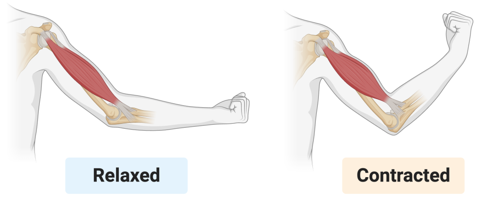
Muscle tissue is able to contract. That’s how we move our arms, or our entire bodies. It’s how we throw balls, lift weights, and reposition our bodies to maintain our balance when we’re cycling, skateboarding, skiing, or surfing. Muscles power how the hairs on our body stand up when we’re cold; how we smile, frown, or raise an eyebrow; how we crack an egg; how we chew our food.
Inside our bodies, muscle contraction is how
- Blood flows through our arteries.
- Food moves through our digestive tract
- Air is drawn into and out of the lungs
- Blood pressure and body temperature are regulated
- The uterus contracts during childbirth.
The key questions for this module on muscle tissue are
- How does muscle tissue contract?
- How is muscle contraction controlled?
We’ll begin with skeletal muscle. Later modules will focus on smooth muscle, which moves materials through organs like the digestive tract and blood vessels, and cardiac muscle, which drives the heartbeat.
2. Muscle: Structure and Connective Tissue Layers
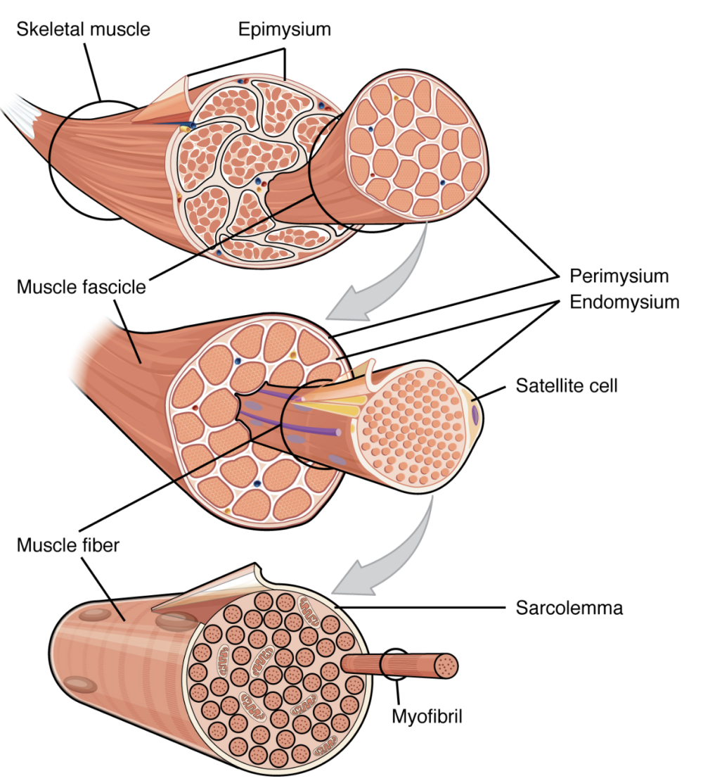
The ability of muscle tissue to contract is rooted in the arrangement of highly organized arrays of proteins found in myofibrils. Myofibrils are long intracellular fibers found within muscle fibers (also known as muscle cells). Let’s start by working our way down from an entire muscle to myofibrils.
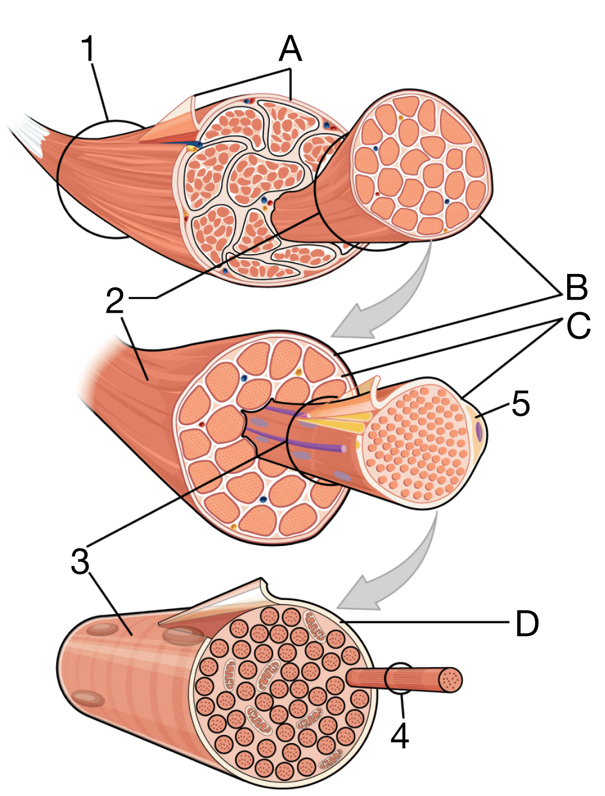
In the diagram on the left, 1 is the entire muscle. You can imagine this being the biceps muscle shown above, which is responsible for bending your arm.
The entire muscle is covered by a thick layer of connective tissue called the epimysium (“A”).
The next level down is the muscle fascicle (“2”). A fascicle is a bundle of muscle fibers, surrounded by connective tissue called perimysium (“B “). How many fascicles are in a typical muscle? There’s no fixed number. For example, what we call the biceps is actually two muscles: the long head and the short head of the biceps brachii. Each head contains dozens to hundreds of fascicles, depending on the size of the muscle and the individual.
Each fascicle consists of numerous muscle fibers (“3”). Muscle fibers are multinucleate cells, meaning they contain multiple nuclei. These nuclei result from muscle development, in which individual precursor cells fuse together to form a single elongated cell with one cell membrane and many nuclei. The membrane is called the sarcolemma (“D”). The many nuclei enable muscle cells to produce the large amount of protein required for contraction.
Muscle fibers are surrounded by a layer of thin connective tissue: the endomysium (“D”).
A muscle fiber contains many myofibrils (“4”), which are long, cylindrical structures responsible for contraction. We’ll examine their molecular structure in more detail below.
The final structure shown in this diagram is 5, a satellite cell. Satellite cells are stem cells that play a key role in muscle growth and repair.
3. Muscle Structure and Connective Tissue Layers: Key
The key below covers everything you read about above. When you’re ready, proceed to the quizzes below.
| Letter | Name | Function |
|---|---|---|
| 1 | Skeletal muscle | Whole muscle organ composed of multiple muscle fascicles that contracts to produce movement. |
| 2 | Muscle fascicle | Bundle of muscle fibers grouped together within a skeletal muscle. |
| 3 | Muscle fiber | Multinucleated muscle cell that contains myofibrils and contracts in response to stimulation. |
| 4 | Myofibril | Rod-like contractile structure within a muscle fiber composed of repeating sarcomeres (the functional units of muscle contraction). |
| 5 | Satellite cell | Stem cell involved in muscle growth, repair, and regeneration. |
| A | Epimysium | Dense connective tissue layer that surrounds and protects the entire skeletal muscle. |
| B | Perimysium | Connective tissue layer that surrounds each muscle fascicle. |
| C | Endomysium | Thin connective tissue layer that surrounds individual muscle fibers. |
| D | Sarcolemma | Plasma membrane of a muscle fiber that conducts electrical signals. |
4. Muscle Structure and Connective Tissue Layers Diagram Quiz
[qwiz use_dataset=”Anatomy Diagrams one letter answers|unit:11.Muscle Tissue|topic:11.1.Skeletal Muscle Structure” style=”width: 600px !important; min-height: 450px !important;” random=”true” qrecord_id=”sciencemusicvideosMeister1961-Muscle Structure and Connective Tissue Layers Quiz”]
[h]Muscle Structure and Connective Tissue Layers
[i]
[x]
[restart]
[/qwiz]
5. Muscle Structure and Connective Tissue Click-on Speed Challenge
[qwiz use_dataset=”Anatomy and Physiology Click-on Challenge Dataset|unit:11.Muscle_Tissue|topic:11.1.Skeletal_Muscle_Structure” style=”width: 600px !important; min-height: 450px !important;” random=”true” quiz_timer=”true” qrecord_id=”sciencemusicvideosMeister1961-Muscle Structure and Connective Tissue Layers Click-on Challenge”]
[h]Muscle Structure and Connective Click-on Speed Challenge
[i]Note the timer in the top right. Your goal is accuracy and speed. A good strategy: once through slowly, then additional trials for improvement.
[x]
[restart]
[/qwiz]
Continue to the next tutorial: myofibrils and sarcomeres