Myofibrils and Sarcomeres: The Sliding Filament Model
1. Introduction and Review
To answer the question “how do muscles contract,” we’re taking a deep look at muscle tissue structure.
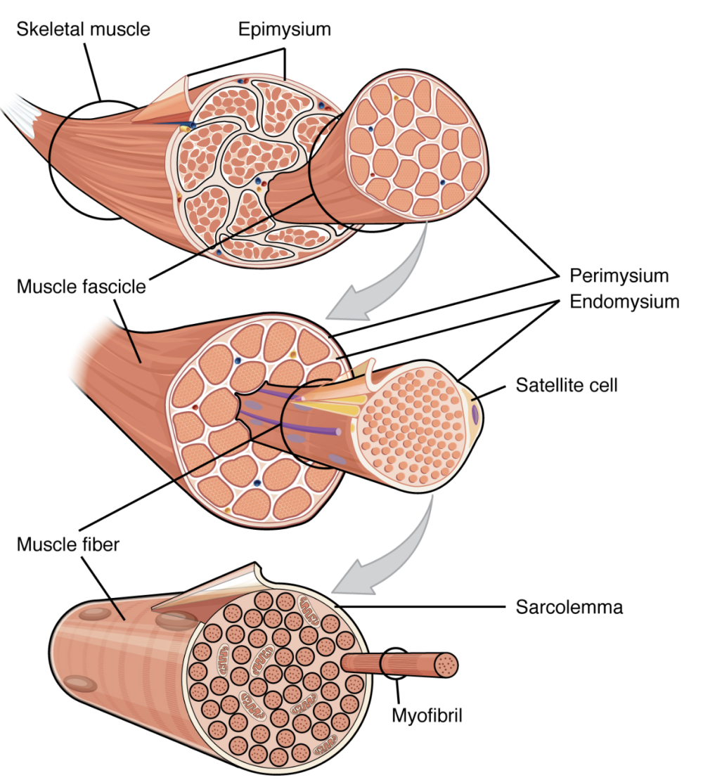
In the previous tutorial, we started that process by learning about the image below. Quickly quiz yourself by identifying the name and function of each indicated structure. Clicking on the diagram will bring up a version with labels.
In this tutorial, we’re going to go all the way down to the level of the two key proteins involved in muscle contraction: actin and myosin. These are organized into a sarcomere: the functional unit of muscle contraction.
2. Myofibrils and Sarcomeres
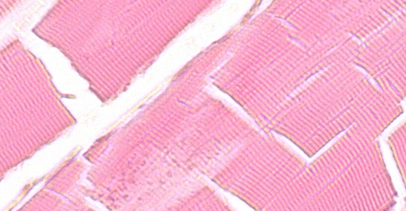
Skeletal muscle is also called striped or striated muscle. The striations are caused by a regularly repeating pattern of molecules within skeletal muscle tissue. Using a light microscope set to high magnification, you can see these striations (though not the underlying molecules). Here’s what it looks like.
To understand these striations, let’s examine the diagram below.
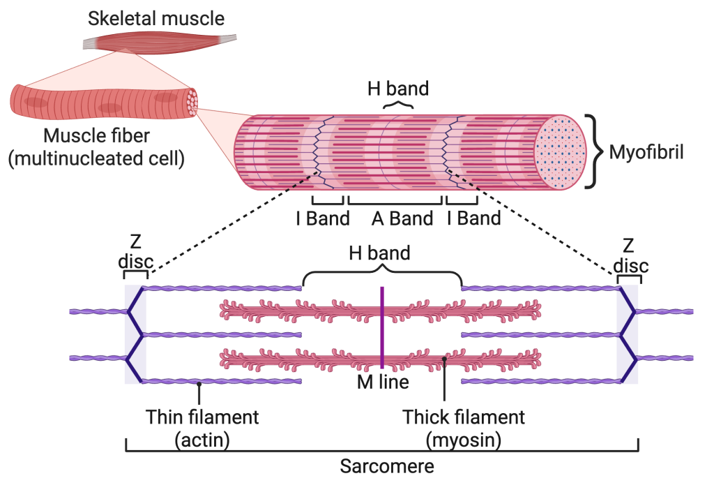
On the top is a skeletal muscle. The first outtake jumps directly to the level of a muscle fiber (a multinucleated cell), where you can see the striated pattern. In the second outtake, which shows a myofibril, the striations are even clearer. The repeating structure, bracketed on the bottom, is called a sarcomere. As we’ll see, sarcomeres are the functional unit of muscle contraction.
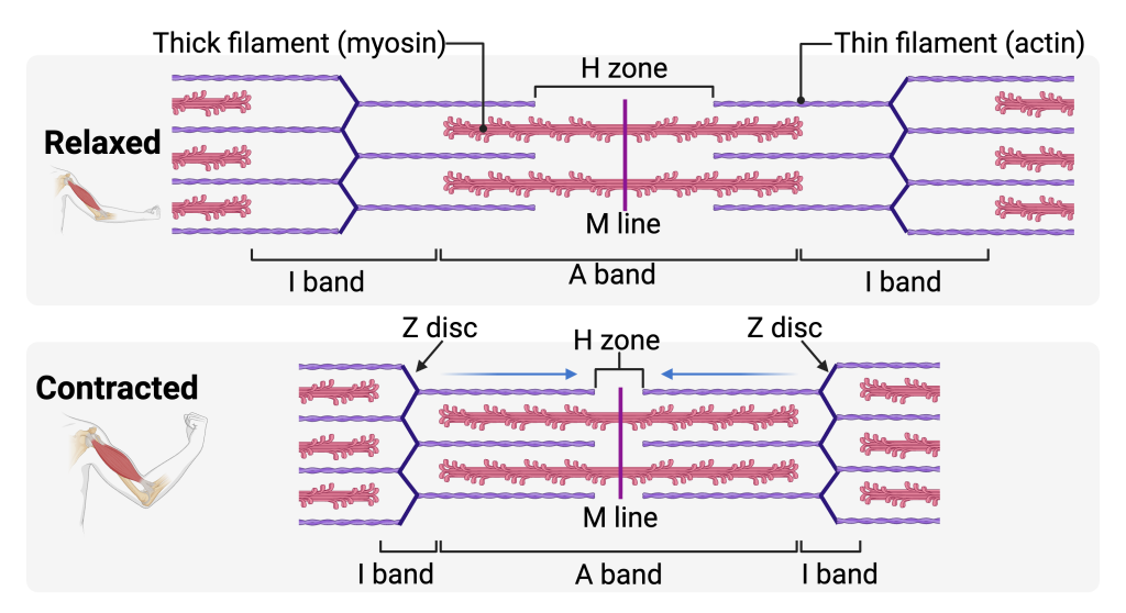
The sarcomere is bracketed by a structure called a Z disc. Z discs are made of proteins that anchor actin filaments (also known as thin filaments). Interspersed between the actin filaments, at the center of the sarcomere, are myosin filaments (also known as thick filaments).
The area of the sarcomere that consists of only thin (actin) filaments is called the I band. Between the I bands is an A band. The A band is equal to the length of the myosin (thick fibers). The ends of the thin filaments also protrude into the A band. The M line is a protein that keeps all of the thick filaments in alignment during a muscle contraction.
If you’ve noticed that the actin filaments look smooth and the myosin filaments look bumpy, you’re on to something important.
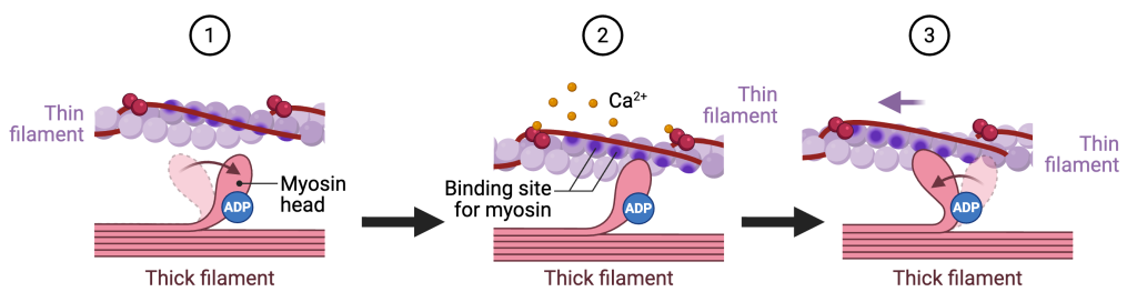
Myosin filaments have “heads” that act as levers to power muscle contraction When a muscle contracts,
- The tip of a myosin molecule (called the “myosin head”) bends forward.
- The myosin head attaches to a myosin-binding site on an actin filament.
- Like a rower pulling back on the oars of a boat, the myosin head pulls back on the actin filament, which has the effect of shortening the sarcomere.
This is called a “power stroke.” In the same way that rowing requires a lot of energy, so does the power stroke: skeletal muscle (which is what you use for rowing) is the largest consumer of ATP in the body.
3. Sarcomeres and Muscle Contraction
 So, what happens during a muscle contraction?
A muscle contracts when its sarcomeres shorten.
In the relaxed state (see the upper image in diagram to your right), the thin (actin) filaments extend partway into the region occupied by the thick (myosin) filaments. The Z discs are farther apart, and the I bands (regions containing only thin filaments) are relatively wide. The H zone, the area that designates where there are thick filaments without overlapping thin filaments, is also relatively wide.
During contraction, myosin heads bind to actin and pull the thin filaments inward, toward the center of the sarcomere (the M line). As this happens:
- The Z discs move closer together
- The I bands become shorter
- The H Zone becomes shorter
- The overlap between actin and myosin increases
Importantly, the filaments themselves do not shorten.
- The thick filaments stay the same length
- The thin filaments stay the same length
- The A band remains constant
What changes is their position. The sliding of thin filaments past thick filaments shortens each sarcomere. When millions of sarcomeres shorten at the same time, myofibrils shorten, muscle fibers shorten, and the entire muscle contracts, producing movement.
This mechanism is called the sliding filament model of muscle contraction. Every time you take a step, throw a ball, or pick up a spoon, that’s what’s happening.
4. The Sarcomere: Checking Understanding
[qwiz style=”min-height: 450px !important; width: 700px !important;” qrecord_id=”sciencemusicvideosMeister1961-Sarcomere Labeled Diagrams”]
[h] The Sarcomere: Checking Understanding
[i]
[q labels=”top”]
[f*] Excellent!
[fx] Sorry, no.
[f*] Correct!
[fx] Sorry, no.
[f*] Correct!
[fx] No, that’s not correct.
[l]Actin
[fx] No. Please try again.
[f*] Good!
[l]Myosin
[fx] No, that’s not correct. Please try again.
[f*] Correct!
[l]Z disk
[fx] No, that’s not correct. Please try again.
[f*] Great!
[l]I band
[fx] No. Please try again.
[f*] Correct!
[l]M line
[fx] No, that’s not correct. Please try again.
[f*] Excellent!
[l]Muscle fiber
[fx] No, that’s not correct. Please try again.
[f*] Good!
[q labels = “top”]
[l]A band
[fx] No, that’s not correct. Please try again.
[f*] Great!
[l]H zone
[fx] No, that’s not correct. Please try again.
[f*] Good!
[l]I band
[fx] No, that’s not correct. Please try again.
[f*] Good!
[l]M line
[fx] No. Please try again.
[f*] Great!
[l]thick filament
[fx] No, that’s not correct. Please try again.
[f*] Excellent!
[l]thin filament
[fx] No. Please try again.
[f*] Good!
[l]Z disc
[fx] No. Please try again.
[f*] Great!
[x]
[restart]
[/qwiz]
5. Another view: Muscle Fibers, Myofibrils, and Sarcomeres
The image below will help you pull together what you’ve learned about muscle tissue so far.
“J” indicates an entire muscle fiber: a multinucleated cell. One of the nuclei is shown at “I,” and mitochondria are shown at “A.” The lighter striations at “H” represent the I bands, which contain only thin (actin) filaments. The darker areas at “G” are the A bands, where thick (myosin) filaments are present (with some portion of the actin fibers overlapping). The membrane of the cell (known as the sarcolemma) is represented by “B.”
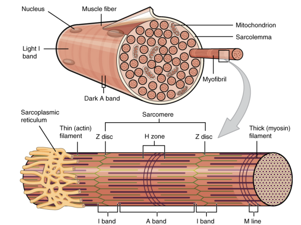
“C” is a single myofibril. “1” represents one sarcomere.
“2” shows the sarcoplasmic reticulum: a specialized form of endoplasmic reticulum that stores and releases the calcium ions that stimulate a sarcomere to contract. That’s a topic that we’ll be addressing in the next tutorial. Actin (thin) filaments are shown at “3,” and the Z disc is represented by “4.”
“5” shows the H Zone. In a relaxed sarcomere, the H-zone contains only thick (myosin) fibers. When a sarcomere contracts, the H zone decreases in length, and might disappear entirely during a complete contraction. “6” points to a thick (myosin) filament.
On the bottom of the myofibril, letter “E” shows the I band, “F” shows the A band, and “D” represents the “M” line (proteins that connect the thick myosin filaments and keep them aligned during muscle contractions).
Once you feel comfortable with the diagram to your left and the ones above, take the quiz below.
| Letter or Number | Name | Function |
|---|---|---|
| A | Mitochondrion | Produces ATP needed to power muscle contraction. |
| B | Sarcolemma | Plasma membrane of the muscle fiber. During contraction, the sarcolemma conducts electrical signals. |
| C | Myofibril | Long sub-cellular fiber composed of repeating sarcomeres. |
| D | M line | Central region of the sarcomere where thick filaments are connected to one another. |
| E | I band (in the myofibril) | Region containing thin filaments only; shortens during contraction. |
| F | A band (in the myofibril) | Region containing thick filaments; length remains constant during contraction. |
| G | A band (on the muscle fiber surface) | See above. It’s the dark band visible on the muscle fiber. |
| H | I band (on the muscle fiber surface) | Light band visible on the muscle fiber where only thin filaments are present. |
| I | Nucleus | Contains genetic information and controls protein synthesis in the muscle fiber. |
| J | Muscle fiber | Multinucleated muscle cell specialized for contraction. |
| 1 | Sarcomere | Functional unit of muscle contraction, extending from Z disc to Z disc. |
| 2 | Sarcoplasmic reticulum | Specialized endoplasmic reticulum of a muscle cell. Stores and releases calcium ions that regulate muscle contraction. |
| 3 | Thin (actin) filament | Anchored to the Z disc and slides during contraction. |
| 4 | Z disc | Defines the boundary of a sarcomere and anchors thin filaments. |
| 5 | H zone | Central region of the A band that, in a relaxed muscle, contains only thick filaments and narrows or disappears during contraction. |
| 6 | Thick (myosin) filament | Contains myosin heads that form cross-bridges with actin. |
6. Myofibril and Sarcomere Structure Quizzes
6a. Quiz 1: Understanding Diagrams
[qwiz use_dataset=”Anatomy Diagrams one letter answers|unit:11.Muscle Tissue|topic:11.2.Muscle Fiber, Myofibrils, and Sarcomeres” style=”width: 600px !important; min-height: 450px !important;” random=”true” qrecord_id=”sciencemusicvideosMeister1961-Myofibril and Sarcomere Structure Diagram Quiz”]
[h]Myofibrils and Sarcomeres
[i]
[x]
[restart]
[/qwiz]
6b. Quiz 2: Understanding Concepts (Multiple Choice)
[qwiz style=”width: 600px !important; min-height: 450px !important;” random=”true” qrecord_id=”sciencemusicvideosMeister1961-Sarcomere Structure MC Quiz”]
[h]Myofibrils and Sarcomeres: Multiple choice
[i]
[q json=”true”] Of the parts of the sarcomere listed below, which part gets shorter when a sarcomere contracts?
[c]IEEgYmFuZA==[Qq]
[f]IE5vLiBUaGUgQSBiYW5kIGNvcnJlc3BvbmRzIHRvIHRoZSBsZW5ndGggb2YgdGhlIHRoaWNrIGZpbGFtZW50cy4gU3R1ZHkgdGhlIGRpYWdyYW0gYmVsb3cgKGFuZCBub3RlIHRoYXQgdGhlIEggem9uZSB3YXMgbm90IG9uZSBvZiB0aGUgY2hvaWNlcyBpbiB0aGlzIHF1ZXN0aW9uKS4=
Cg==[Qq]
[c]IEkgYm FuZA==[Qq]
[f]IEV4Y2VsbGVudCEgQXMgeW91IGNhbiBzZWUgYmVsb3csIHdoZW4gYSBzYXJjb21lcmUgY29udHJhY3RzLCB0aGUgSSBiYW5kIGRlY3JlYXNlcyBpbiBsZW5ndGguIE5vdGUgdGhhdCB0aGUgSCB6b25lIGFsc28gZGVjcmVhc2VzIChidXQgdGhhdCB3YXNuJiM4MjE3O3Qgb25lIG9mIHRoZSBjaG9pY2VzIGluIHRoaXMgcXVlc3Rpb24pLg==
Cg==[Qq]
[c]IE0gbGluZQ==[Qq]
[f]IE5vLiBUaGUgTSBsaW5lIGhlbHBzIG9yZ2FuaXplIHRoaWNrIGZpbGFtZW50cy7CoFN0dWR5IHRoZSBkaWFncmFtIGJlbG93IChhbmQgbm90ZSB0aGF0IHRoZSBIIHpvbmUgd2FzIG5vdCBvbmUgb2YgdGhlIGNob2ljZXMgaW4gdGhpcyBxdWVzdGlvbiku
Cg==[Qq]
[c]IFogZGlzYw==[Qq]
[f]IE5vLiBaIGRpc2NzIG1vdmUgY2xvc2VyIHRvZ2V0aGVyIGR1cmluZyBjb250cmFjdGlvbiwgYnV0IHRoZWlyIHNpemUgZG9lcyBub3QgY2hhbmdlLiBTdHVkeSB0aGUgZGlhZ3JhbSBiZWxvdyAoYW5kIG5vdGUgdGhhdCB0aGUgSCB6b25lIHdhcyBub3Qgb25lIG9mIHRoZSBjaG9pY2VzIGluIHRoaXMgcXVlc3Rpb24pLg==
Cg==[Qq]
[q json=”true”] Which statement best describes what happens to actin and myosin filaments during muscle contraction?
[c]IEJvdGggYWN0aW4gYW5kIG15b3NpbiBmaWxhbWVudHMgc2hvcnRlbg==[Qq]
[f]IE5vLiBTdHVkeSB0aGUgZGlhZ3JhbSBiZWxvdy4gVGhpbmsgY2FyZWZ1bGx5IGFib3V0IHdoZXRoZXIgdGhlIGZpbGFtZW50cyB0aGVtc2VsdmVzIGNoYW5nZSBsZW5ndGggb3Igd2hldGhlciB0aGVpciByZWxhdGl2ZSBwb3NpdGlvbiBjaGFuZ2VzLg==
Cg==[Qq]
[c]IEFjdGluIGZpbGFtZW50cyBzbGlkZS BwYXN0IG15b3NpbiBmaWxhbWVudHM=[Qq]
[f]IENvcnJlY3QhIEFzIHlvdSBjYW4gc2VlIGJlbG93LCBtdXNjbGUgY29udHJhY3Rpb24gb2NjdXJzIHdoZW4gdGhpbiBmaWxhbWVudHMgc2xpZGUgaW53YXJkIHBhc3QgdGhpY2sgZmlsYW1lbnRzLg==
Cg==[Qq]
[c]IE15b3NpbiBmaWxhbWVudHMgc2xpZGUgcGFzdCBhY3RpbiBmaWxhbWVudHMgYW5kIHNob3J0ZW4=[Qq]
[f]IE5vLiBNeW9zaW4gZmlsYW1lbnRzIGRvIG1vdmUgcmVsYXRpdmUgdG8gYWN0aW4sIGJ1dCB0aGVpciBsZW5ndGggZG9lcyBub3QgY2hhbmdlLiBTdHVkeSB0aGUgZGlhZ3JhbSBiZWxvdyBhbmQgc2VlIGlmIHlvdSBjYW4gaWRlbnRpZnkgdGhlIGNvcnJlY3QgYW5zd2VyLg==
Cg==[Qq]
[c]IEFjdGluIGZpbGFtZW50cyBiZW5kIHdoaWxlIG15b3NpbiBmaWxhbWVudHMgcmVtYWluIHN0YXRpb25hcnk=[Qq]
[f]IE5vLiBOZWl0aGVyIGZpbGFtZW50IGJlbmRzLiBTdHVkeSB0aGUgZGlhZ3JhbSBiZWxvdyBhbmQgc2VlIGlmIHlvdSBjYW4gaWRlbnRpZnkgdGhlIGNvcnJlY3QgYW5zd2VyLg==
Cg==
Cg==[Qq]
[q json=”true”] Why are sarcomeres described as the functional unit of muscle contraction?
[c]IFRoZXkgZ2VuZXJhdGUgQVRQIG5lZWRlZCBmb3IgY29udHJhY3Rpb24=[Qq]
[f]IE5vLiBBVFAgaXMgZXNzZW50aWFsLCBidXQgaXQgaXMgcHJvZHVjZWQgYnkgbWl0b2Nob25kcmlhLCBub3QgYnkgc2FyY29tZXJlcy4gVGhpbmsgYWJvdXQgd2hpY2ggc3RydWN0dXJlIGFjdHVhbGx5IHNob3J0ZW5zIHRvIHByb2R1Y2UgZm9yY2Uu[Qq]
[c]IFRoZXkgdHJhbnNtaXQgbmVydmUgaW1wdWxzZXMgdG8gbXVzY2xlIGZpYmVycw==[Qq]
[f]IE5vLiBTYXJjb21lcmVzIG1ha2UgdXAgbXlvZmlicmlscywgd2hpY2ggbWFrZSB1cCBtdXNjbGUgZmliZXJzLiBTdHVkeSB0aGUgZGlhZ3JhbSBiZWxvdyBhbmQgc2VlIGlmIHlvdSBjYW4gaWRlbnRpZnkgdGhlIGNvcnJlY3QgYW5zd2VyLg==
Cg==[Qq]
[c]IFRoZXkgc2hvcnRlbiBkdXJpbmcgY29udH JhY3Rpb24gYW5kIGdlbmVyYXRlIGZvcmNl[Qq]
[f]IENvcnJlY3QhIEFzIHNob3duIGJlbG93LCBzYXJjb21lcmVzIHNob3J0ZW4gYXMgYWN0aW4gc2xpZGVzIHBhc3QgbXlvc2luLCBwcm9kdWNpbmcgZm9yY2Uu
Cg==[Qq]
[c]IFRoZXkgc3RvcmUgY2FsY2l1bSBpb25zIHVzZWQgZHVyaW5nIGNvbnRyYWN0aW9u[Qq]
[f]IE5vLiBDYWxjaXVtIGlzIGNyaXRpY2FsIGZvciBtdXNjbGUgY29udHJhY3Rpb24sIGJ1dCBpdCBpcyBzdG9yZWQgaW4gdGhlIHNhcmNvcGxhc21pYyByZXRpY3VsdW0uIFN0dWR5IHRoZSBkaWFncmFtIGJlbG93IGFuZCBzZWUgaWYgeW91IGNhbiBpZGVudGlmeSB0aGUgY29ycmVjdCBhbnN3ZXIu
Cg==[Qq]
[q json=”true”] Which structure anchors the thin (actin) filaments in a sarcomere?
[c]IE0gbGluZQ==[Qq]
[f]IE5vLiBUaGUgTSBsaW5lIGlzIGFzc29jaWF0ZWQgd2l0aCB0aGljayBmaWxhbWVudHMuIFN0dWR5IHRoZSBkaWFncmFtIGJlbG93IGFuZCBzZWUgaWYgeW91IGNhbiBpZGVudGlmeSB3aGljaCBzdHJ1Y3R1cmUgaXMgYW5jaG9yaW5nIHRoZSB0aGluIGZpbGFtZW50cy4=
Cg==[Qq]
[c]IFogZG lzYw==[Qq]
[f]IEV4Y2VsbGVudCEgWiBkaXNjcyBhbmNob3IgdGhpbiBmaWxhbWVudHMgYW5kIGRlZmluZSB0aGUgYm91bmRhcmllcyBvZiBlYWNoIHNhcmNvbWVyZS4=
Cg==[Qq]
[c]IEEgYmFuZA==[Qq]
[f]IE5vLiBUaGUgQSBiYW5kIGlzIGEgcmVnaW9uLCBub3QgYW4gYW5jaG9yaW5nIHN0cnVjdHVyZS4gU3R1ZHkgdGhlIGRpYWdyYW0gYmVsb3cgYW5kIHNlZSBpZiB5b3UgY2FuIGlkZW50aWZ5IHdoaWNoIHN0cnVjdHVyZSBpcyBhbmNob3JpbmcgdGhlIHRoaW4gZmlsYW1lbnRzLg==
Cg==[Qq]
[c]IEggem9uZSAobGFiZWxlZCBhcyB0aGUgSCBiYW5kIGluIHRoZSBkaWFncmFtKQ==[Qq]
[f]IE5vLiBUaGUgSCB6b25lIGlzIGRlZmluZWQgYnkgZmlsYW1lbnQgYXJyYW5nZW1lbnQsIG5vdCBhdHRhY2htZW50LiBTdHVkeSB0aGUgZGlhZ3JhbSBiZWxvdyBhbmQgc2VlIGlmIHlvdSBjYW4gaWRlbnRpZnkgd2hpY2ggc3RydWN0dXJlIGlzIGFuY2hvcmluZyB0aGUgdGhpbiBmaWxhbWVudHMu
Cg==[Qq]
[q json=”true”] Which region of a sarcomere contains only thin filaments in a relaxed muscle?
[c]IEEgYmFuZA==[Qq]
[f]IE5vLiBUaGUgQSBiYW5kIGNvbnRhaW5zIHRoaWNrIGZpbGFtZW50cy4gTG9vayBmb3IgYSByZWdpb24gd2hlcmUgbXlvc2luIGlzIGFic2VudC4=
Cg==[Qq]
[c]IEkgYm FuZA==[Qq]
[f]IENvcnJlY3QhIFRoZSBJIGJhbmQgY29udGFpbnMgb25seSB0aGluIGZpbGFtZW50cyBpbiBhIHJlbGF4ZWQgc2FyY29tZXJlLg==
Cg==[Qq]
[c]IEggem9uZQ==[Qq]
[f]IE5vLiBUaGUgSCB6b25lIGNvbnRhaW5zIG9ubHkgdGhpY2sgZmlsYW1lbnRzLiBMb29rIGZvciBhIHJlZ2lvbiB3aGVyZSBteW9zaW4gaXMgYWJzZW50Lg==
Cg==[Qq]
[c]IE0gbGluZQ==[Qq]
[f]IE5vLiBUaGUgTSBsaW5lIGlzIGEgcHJvdGVpbiBzdHJ1Y3R1cmUgdGhhdCBrZWVwcyB0aGUgdGhpY2sgZmlsYW1lbnRzIGluIGFsaWdubWVudCBkdXJpbmcgYSBtdXNjbGUgY29udHJhY3Rpb24uIExvb2sgZm9yIGEgcmVnaW9uIHdoZXJlIG15b3NpbiBpcyBhYnNlbnQu
Cg==[Qq]
[q json=”true”] Which statement about the A band is correct?
[c]IEl0IGJlY29tZXMgc2hvcnRlciBkdXJpbmcgY29udHJhY3Rpb24=[Qq]
[f]IE5vLiBTdHVkeSB0aGUgZGlhZ3JhbSBiZWxvdy4gRG9lcyB0aGUgbGVuZ3RoIG9mIHRoZSBBIGJhbmQgY2hhbmdlIGR1cmluZyBjb250cmFjdGlvbj8=
Cg==[Qq]
[c]IEl0cyBsZW5ndGggcmVtYWlucyBjb25z dGFudCBkdXJpbmcgY29udHJhY3Rpb24=[Qq]
[f]IENvcnJlY3QhIFRoZSBBIGJhbmQgc3RheXMgdGhlIHNhbWUgbGVuZ3RoIGJlY2F1c2UgdGhpY2sgZmlsYW1lbnRzIGRvIG5vdCBzaG9ydGVuLg==
Cg==[Qq]
[c]IEl0IGNvbnRhaW5zIG9ubHkgdGhpbiBmaWxhbWVudHM=[Qq]
[f]IE5vLiBUaGUgQSBiYW5kIGlzIGRlZmluZWQgYnkgdGhlIHByZXNlbmNlIG9mIHRoaWNrIGZpbGFtZW50cy4gU3R1ZHkgdGhlIGRpYWdyYW0gYmVsb3cgYW5kIHRyeSB0byBpZGVudGlmeSB0aGUgY29ycmVjdCBhbnN3ZXIu
Cg==[Qq]
[c]IEl0IGRpc2FwcGVhcnMgZHVyaW5nIGNvbnRyYWN0aW9u[Qq]
[f]IE5vLiBEdXJpbmcgbXVzY2xlIGNvbnRyYWN0aW9uLCB0aGUgZmlsYW1lbnRzIHNsaWRlIHBhc3Qgb25lIGFub3RoZXIsIHNob3J0ZW5pbmcgdGhlIHNhcmNvbWVyZSB3aXRob3V0IGNoYW5naW5nIGxlbmd0aC4gU3R1ZHkgdGhlIGRpYWdyYW0gYmVsb3cgYW5kIHRyeSB0byBpZGVudGlmeSB0aGUgY29ycmVjdCBhbnN3ZXIu
Cg==[Qq]
[q json=”true”] What causes the striated (striped) appearance of skeletal muscle?
[c]IFRoZSBwcmVzZW5jZSBvZiBtdWx0aXBsZSBudWNsZWkgaW4gbXVzY2xlIGZpYmVycw==[Qq]
[f]IE5vLiBTdHVkeSB0aGUgZGlhZ3JhbSBiZWxvdy4gTnVjbGVpIGFyZSBpbXBvcnRhbnQgZm9yIHByb3RlaW4gc3ludGhlc2lzLCBidXQgdGhleSBkbyBub3QgY3JlYXRlIGEgcmVwZWF0aW5nIHBhdHRlcm4uIFdoYXQgZG9lcz8=
Cg==[Qq]
[c]IFRoZSByZWd1bGFyIGFycmFuZ2VtZW50IG9mIGFjdGluIGFu ZCBteW9zaW4gaW50byByZXBlYXRpbmcgc2FyY29tZXJlcw==[Qq]
[f]IENvcnJlY3QhIFRoZSByZXBlYXRpbmcgcGF0dGVybiBvZiBBIGJhbmRzIGFuZCBJIGJhbmRzIGNyZWF0ZXMgdmlzaWJsZSBzdHJpYXRpb25zLg==
Cg==[Qq]
[c]IEZvbGRpbmcgb2YgdGhlIHNhcmNvbGVtbWE=[Qq]
[f]IE5vLiBTdHVkeSB0aGUgZGlhZ3JhbSBiZWxvdyBhbmQgc2VlIGlmIHlvdSBjYW4gaWRlbnRpZnkgdGhlIHJlcGVhdGluZyBzdHJ1Y3R1cmUgdGhhdCBjYXVzZXMgc3RyaWF0aW9ucy4=
Cg==[Qq]
[c]IERpZmZlcmVuY2VzIGluIG1pdG9jaG9uZHJpYWwgZGVuc2l0eQ==[Qq]
[f]IE5vLiBTdHVkeSB0aGUgZGlhZ3JhbSBiZWxvdyBhbmQgc2VlIGlmIHlvdSBjYW4gaWRlbnRpZnkgdGhlIHJlcGVhdGluZyBzdHJ1Y3R1cmUgdGhhdCBjYXVzZXMgc3RyaWF0aW9ucy4=
Cg==[Qq]
[q json=”true”] Which event directly causes a sarcomere to shorten?
[c]IFRoaWNrIGZpbGFtZW50cyBtb3ZpbmcgdG93YXJkIHRoZSBaIGRpc2Nz[Qq]
[f]IE5vLiBUaGljayBmaWxhbWVudHMgcmVtYWluIGNlbnRlcmVkLiBTdHVkeSB0aGUgZGlhZ3JhbSBiZWxvdyBhbmQgc2VlIGlmIHlvdSBjYW4gaWRlbnRpZnkgdGhlIGNvcnJlY3QgYW5zd2VyLg==
Cg==[Qq]
[c]IFRoaW4gZmlsYW1lbnRzIHNsaWRp bmcgdG93YXJkIHRoZSBNIGxpbmU=[Qq]
[f]IENvcnJlY3QhIFRoaW4gZmlsYW1lbnRzIHNsaWRlIGlud2FyZCB0b3dhcmQgdGhlIGNlbnRlciBvZiB0aGUgc2FyY29tZXJlLCBzaG9ydGVuaW5nIGl0Lg==
Cg==[Qq]
[c]IFogZGlzY3MgcHVsbGluZyBvbiBhY3RpbiBmaWxhbWVudHM=[Qq]
[f]IE5vLiBaIGRpc2NzIG1vdmUgYXMgYSByZXN1bHQgb2YgY29udHJhY3Rpb24sIG5vdCBhcyB0aGUgY2F1c2UuIFN0dWR5IHRoZSBkaWFncmFtIGJlbG93IGFuZCBzZWUgaWYgeW91IGNhbiBpZGVudGlmeSB0aGUgY29ycmVjdCBhbnN3ZXIu
Cg==[Qq]
[c]IE15b3NpbiBmaWxhbWVudHMgc2hvcnRlbmluZw==[Qq]
[f]IE5vLiBGaWxhbWVudCBsZW5ndGggZG9lcyBub3QgY2hhbmdlLiBTdHVkeSB0aGUgZGlhZ3JhbSBiZWxvdyBhbmQgc2VlIGlmIHlvdSBjYW4gaWRlbnRpZnkgdGhlIGNvcnJlY3QgYW5zd2VyLg==
Cg==[Qq]
[q json=”true”] Which structure helps keep thick filaments aligned during contraction?
[c]IFogZGlzYw==[Qq]
[f]IE5vLiBaIGRpc2NzIGFuY2hvciB0aGluIGZpbGFtZW50cy4gU3R1ZHkgdGhlIGRpYWdyYW0gYmVsb3cuIFdoYXQgY291bGQga2VlcCB0aGUgdGhpY2sgZmlsYW1lbnRzIGFsaWduZWQ/
Cg==[Qq]
[c]IE0gbG luZQ==[Qq]
[f]IENvcnJlY3QhIFRoZSBNIGxpbmUgYW5jaG9ycyBhbmQgYWxpZ25zIHRoaWNrIGZpbGFtZW50cyBkdXJpbmcgY29udHJhY3Rpb24u
Cg==[Qq]
[c]IEkgYmFuZA==[Qq]
[f]IE5vLiBUaGUgSSBiYW5kIGlzIGEgcmVnaW9uLCBub3QgYW4gYW5jaG9yaW5nIHN0cnVjdHVyZS4gU3R1ZHkgdGhlIGRpYWdyYW0gYmVsb3cuIFdoYXQgY291bGQga2VlcCB0aGUgdGhpY2sgZmlsYW1lbnRzIGFsaWduZWQ/
Cg==[Qq]
[c]IFNhcmNvbWVyZQ==[Qq]
[f]IE5vLiBUaGUgc2FyY29tZXJlIGlzIHRoZSBlbnRpcmUgZnVuY3Rpb25hbCB1bml0IG9mIGNvbnRyYWN0aW9uIHdpdGhpbiBtdXNjbGUgdGlzc3VlLiBTdHVkeSB0aGUgZGlhZ3JhbSBiZWxvdy4gV2hhdCBjb3VsZCBrZWVwIHRoZSB0aGljayBmaWxhbWVudHMgYWxpZ25lZD8=
Cg==[Qq]
[q json=”true”] During contraction, which of the following changes occurs?
[c]IFRoaWNrIGZpbGFtZW50cyBiZWNvbWUgc2hvcnRlcg==[Qq]
[f]IE5vLiBGaWxhbWVudCBsZW5ndGggcmVtYWlucyBjb25zdGFudC4gU3R1ZHkgdGhlIGRpYWdyYW0gYmVsb3cgYW5kIHNlZSBpZiB5b3UgY2FuIGlkZW50aWZ5IHRoZSBjb3JyZWN0IGFuc3dlci4=
Cg==[Qq]
[c]IFRoaW4gZmlsYW1lbnRzIGJlY29tZSBzaG9ydGVy[Qq]
[f]IE5vLiBOZWl0aGVyIHRoZSB0aGljayBmaWxhbWVudHMgbm9yIHRoZSB0aGluIGZpbGFtZW50cyBjaGFuZ2UgaW4gbGVuZ3RoIHdoZW4gYSBtdXNjbGUgY29udHJhY3RzLiBTdHVkeSB0aGUgZGlhZ3JhbSBiZWxvdyBhbmQgc2VlIGlmIHlvdSBjYW4gaWRlbnRpZnkgdGhlIGNvcnJlY3QgYW5zd2VyLg==
Cg==[Qq]
[c]IE92ZXJsYXAgYmV0d2VlbiBhY3Rpbi BhbmQgbXlvc2luIGluY3JlYXNlcw==[Qq]
[f]IENvcnJlY3QhIEluY3JlYXNlZCBvdmVybGFwIGJldHdlZW4gZmlsYW1lbnRzIHNob3J0ZW5zIHRoZSBzYXJjb21lcmUu
Cg==[Qq]
[c]IFRoZSBBIGJhbmQgbmFycm93cw==[Qq]
[f]IE5vLiBUaGUgQSBiYW5kIHJlbWFpbnMgdGhlIHNhbWUgbGVuZ3RoLiBTdHVkeSB0aGUgZGlhZ3JhbSBiZWxvdyBhbmQgc2VlIGlmIHlvdSBjYW4gaWRlbnRpZnkgdGhlIGNvcnJlY3QgYW5zd2VyLg==
Cg==[Qq]
[q json=”true”] What happens to the H zone during a strong muscle contraction?
[c]IEl0IGJlY29tZXMgd2lkZXI=[Qq]
[f]IE5vLiBTdHVkeSB0aGUgZGlhZ3JhbSBiZWxvdyBhbmQgc2VlIGlmIHlvdSBjYW4gaWRlbnRpZnkgdGhlIGNvcnJlY3QgYW5zd2VyLg==
Cg==[Qq]
[c]IEl0IG5hcnJvd3Mgb3Ig bWF5IGRpc2FwcGVhcg==[Qq]
[f]IENvcnJlY3QhIEFzIG92ZXJsYXAgaW5jcmVhc2VzLCB0aGUgSCB6b25lIGRlY3JlYXNlcyBpbiBzaXplIGFuZCBtYXkgZGlzYXBwZWFyLg==
Cg==[Qq]
[c]IEl0IHR1cm5zIGludG8gYW4gSSBiYW5k[Qq]
[f]IE5vLiBTdHVkeSB0aGUgZGlhZ3JhbSBiZWxvdy4gRm9jdXMgb24gdGhlIEggem9uZSBhbmQgc2VlIGlmIHlvdSBjYW4gaWRlbnRpZnkgdGhlIGNvcnJlY3QgYW5zd2VyLg==
Cg==[Qq]
[c]IEl0IHJlbWFpbnMgdW5jaGFuZ2Vk[Qq]
[f]IE5vLiBTdHVkeSB0aGUgZGlhZ3JhbSBiZWxvdy4gRm9jdXMgb24gdGhlIEggem9uZSBhbmQgc2VlIGlmIHlvdSBjYW4gaWRlbnRpZnkgdGhlIGNvcnJlY3QgYW5zd2VyLg==
Cg==[Qq]
[x]
[restart]
[/qwiz]
Continue to the next tutorial: Control of Muscle Contraction