Control of Muscle Contraction, Part 1: Neuromuscular Junction
1. Introduction and Review
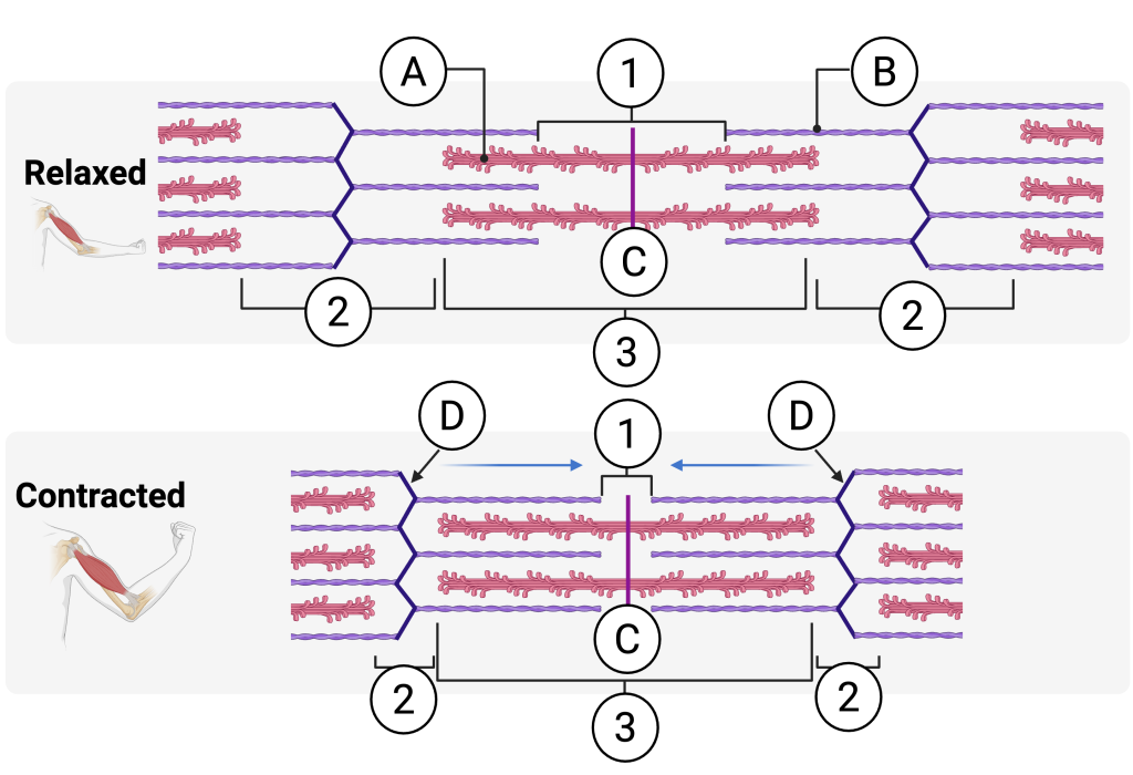
In the previous tutorial, you learned about the molecular basis of muscle contraction. To review, quiz yourself on the diagram below.
However, we’re still left with a question. How does the body control when a muscle contracts? This is especially evident with skeletal muscles, which are voluntary. That means you think a thought, and your muscles respond. Everything you’ve ever accomplished — from each step you’ve taken to each keyboard key you’ve clicked — depends on your ability to voluntarily control what your muscles do. How does that work?
2. Neuromuscular Junctions
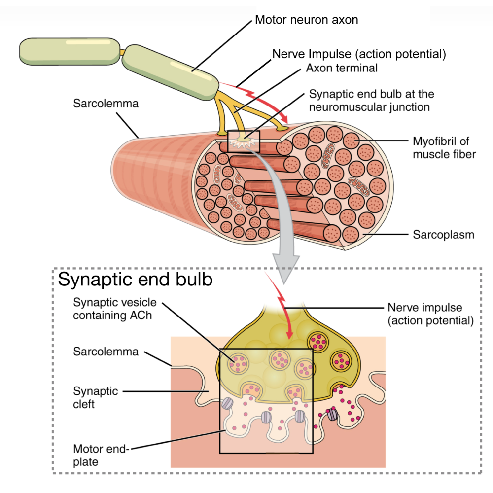
For a thought to be converted into a muscle contraction, a signal needs to be sent from your brain to a muscle. The diagram on the left shows the very end of that process. Letter “B” represents an axon. You can think of axons as long wires that extend from a nerve cell to whatever target that nerve cell connects with. The signal itself — a nerve impulse — is represented by “C.” Letter “D” represents the axon terminal: that’s where the axon ends.
“E” is the synaptic end bulb. It’s a swelling at the end of the axon. The synaptic end bulb is enlarged in the inset at the bottom of the diagram (also labeled “E”). The function of the end bulb is to transmit the nerve impulse (“C”) to the muscle fiber’s membrane: the sarcolemma (“A”).
Nerve impulse transmission is often compared to electrical wiring (an analogy I used above when I compared an axon to a wire). But there’s one big difference. Electrical wires almost always directly touch whatever they supply with electricity. By contrast, nerve cells connect with their targets at a synapse. At the synapse, the end of the nerve cell doesn’t actually touch its target. Instead, it is separated from its target by a small, fluid-filled space. This space is called a synaptic cleft, and it’s represented by letter “I.”
To send a signal across the synaptic cleft, the axon releases a molecule called acetylcholine (ACh) from synaptic vesicles (“J”). Acetylcholine is a neurotransmitter. Its function is to diffuse across the synaptic cleft and to bind with receptors embedded in the motor end plate, the area of muscle fiber membrane that’s directly opposite the synaptic end bulb.
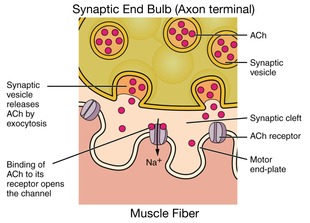
An enlarged version of the structures in the synaptic cleft is shown at right. “I” represents the tip of the axon that’s sending the message. “F” represents the muscle fiber that’s receiving the message. “A” is a molecule of acetylcholine, held within a vesicle (“B”). At “H,” we see release of acetylcholine through exocytosis: one of the vesicles fuses with the membrane at the end of the axon, releasing its contents into the synaptic cleft. The acetylcholine diffuses across the synaptic cleft (“C”) until it binds to a membrane receptor in the sarcolemma (one of which is shown at “D”). The muscle fiber membrane at this position —directly across the synapse from the axonal bulb — is called a motor end plate (“E”). It’s wavy form increases its surface area, allowing for the placement of more receptors.
These receptors are also channels. When acetylcholine binds with the receptor (shown at “G”), the channel opens, allowing a sodium ion (Na+) to diffuse through the sarcolemma and enter the muscle cell.
When enough sodium ions diffuse through the muscle fiber’s membrane, the membrane becomes excited. Excitation, in this context, means that a process gets unleashed that ultimately results in a muscle contraction. The process is called an action potential: a wave of additional sodium ions that quickly diffuse across the muscle cell’s membrane. Because the process involves nerve cell excitation that culminates in muscle contraction, the entire process is called excitation-contraction coupling, the details of which we’ll cover below, and in the next tutorial. But first, take this quiz.
3. Neuromuscular Junction Quiz
[qwiz random=”true” qrecord_id=”sciencemusicvideosMeister1961-Neuromuscular junction: Fill-in-the-Blanks”]
[h]Neuromuscular junction: Fill-in-the-Blanks Quiz
[i]
[q]In the diagram below “A” is the membrane of a nerve cell fiber, also known as the [hangman].
[c]c2FyY29sZW1tYQ==[Qq]
[q]In the diagram below, “B” is the long extension of a nerve cell, also known as an [hangman].
[c]QXhvbg==[Qq]
[q]In the diagram below “C” represents a nerve [hangman].
[c]aW1wdWxzZQ==[Qq]
[q]In the diagram below “D” is the [hangman] [hangman]
[c]YXhvbg==[Qq]
[c]dGVybWluYWw=[Qq]
[q]In the diagram below, “E” represents the [hangman] end bulb.
[c]c3luYXB0aWM=[Qq]
[q]In the diagram below, “F” represents bundles of contractile threads inside the muscle fiber called [hangman].
[c]bXlvZmlicmlscw==[Qq]
[q]In the diagram below, “G” labels the fluid-filled interior of a muscle fiber. In a typical cell, this is known as cytoplasm. But in muscle cells, it’s known as the [hangman].
[c]c2FyY29wbGFzbQ==[Qq]
[q]In the enlarged view below, “H” represents the specialized region of the muscle fiber membrane that contains receptors for acetylcholine, called the motor [hangman]-[hangman].
[c]ZW5k[Qq]
[c]cGxhdGU=[Qq]
[q]In the enlarged view below, “I” labels the small fluid-filled gap between the nerve cell and the muscle fiber, known as the synaptic [hangman].
[c]Y2xlZnQ=[Qq]
[q]In the enlarged view below, “J” represents membrane-bound structures that store acetylcholine before it is released. These are called synaptic [hangman].
[c]dmVzaWNsZXM=[Qq]
[q]In the diagram below, “I” represents the [hangman] end-bulb
[c]c3luYXB0aWM=[Qq]
[q]In the diagram below, “B” represents a synaptic [hangman].
[c]dmVzaWNsZQ==[Qq]
[q]In the diagram below, “A” labels a molecule of [hangman].
[c]YWNldHlsY2hvbGluZQ==[Qq]
[q]In the diagram below, “C” represents the synaptic [hangman].
[c]Y2xlZnQ=[Qq]
[q]In the diagram below, “D” labels an ACh [hangman].
[c]cmVjZXB0b3I=[Qq]
[q]In the diagram below, “E” represents the motor end-[hangman].
[c]cGxhdGU=[Qq]
[q]In the diagram below, “F” represents the membrane of a [hangman] fiber.
[c]bXVzY2xl[Qq]
[q]In the diagram below, “G” shows how when the Ach binds to its [hangman], a sodium [hangman] opens.
[c]cmVjZXB0b3I=[Qq]
[c]Y2hhbm5lbA==[Qq]
[q]In the diagram below, “H” represents [hangman]. That’s when a [hangman] fuses with the membrane and releases its contents (in this case, the neurotransmitter acetylcholine.
[c]ZXhvY3l0b3Npcw==[Qq]
[c]IHZlc2ljbGU=[Qq]
[q]In the diagram below, “a” represents acetylcholine, which is a key [hangman] involved in the stimulation of muscle contraction.
[c]bmV1cm90cmFuc21pdHRlcg==[Qq]
[x]
[restart]
[/qwiz]
4. Excitation-Contraction Coupling
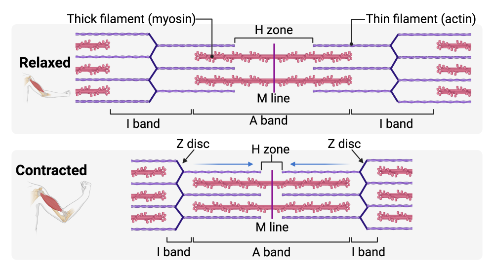
Above, we saw how a nerve impulse can lead to an action potential that moves along the surface of the sarcolemma— the muscle fiber cell membrane. Now let’s see how that impulse will get to the sarcomeres, causing them to contract (as shown at the top of this page).
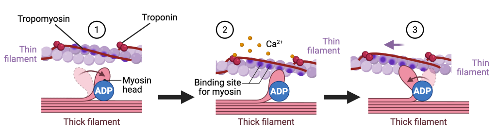
For sarcomeres to contract, the heads of myosin molecules need to bind with myosin-binding sites on actin and pull the actin toward the Z discs. In the relaxed (uncontracted) sarcomere shown in diagram 1, myosin can’t bind with actin because the binding sites are blocked by a protein fiber called tropomyosin. The position of tropomyosin is controlled by another protein called troponin. In diagram 2, calcium ions (Ca²⁺) bind to troponin, which nudges the tropomyosin, exposing the myosin-binding sites, allowing the myosin head to bind. In diagram 3, we see the result: myosin bends, pulling back on the thin filament. That’s the molecular basis of muscle contraction.
That leads to two questions:
- Where does the calcium come from?
- How is its release controlled?
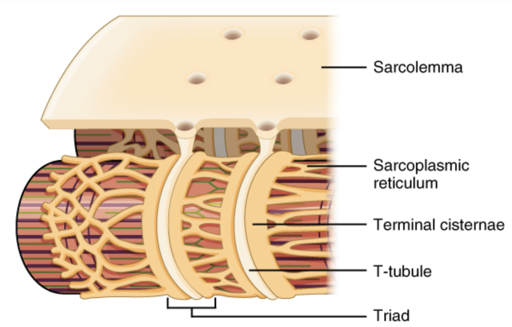
Calcium is stored in the sarcoplasmic reticulum (SR), which forms a network of tubules that surrounds each myofibril. Control of calcium release depends on the layout of the sarcolemma (the muscle cell membrane). The sarcolemma doesn’t just cover the surface of the muscle fiber; it penetrates deep into the sarcoplasm through invaginations called T-tubules.
The T-tubules are flanked by two terminal cisternae: parts of the sarcoplasmic reticulum that form a ring surrounding the myofibril. The combination of T-tubule and their two flanking terminal cisternae is known as a triad. As the action potential moves through the T-tubules, calcium ions (Ca²⁺) are released from the cisternae into the sarcoplasm, enabling the sarcomeres to contract.
5. Quiz: Neuromuscular Junctions
[qwiz random=”true” use_dataset=”Anatomy Diagrams one letter answers|unit:11.Muscle Tissue|topic:11.3.Neuromuscular Junctions” qrecord_id=”sciencemusicvideosMeister1961-Neuromuscular Junction: Diagram Questions”]
[h]Neuromuscular Junction: Diagram Questions
[i]
[/qwiz]
Continue to the next tutorial: ATP and the Cross-Bridge Cycle