Control of Muscle Contraction Part 2: ATP and the Cross Bridge Cycle
1. Introduction
In previous tutorials in this module, we’ve established that
- Muscles contract when their sarcomeres shorten
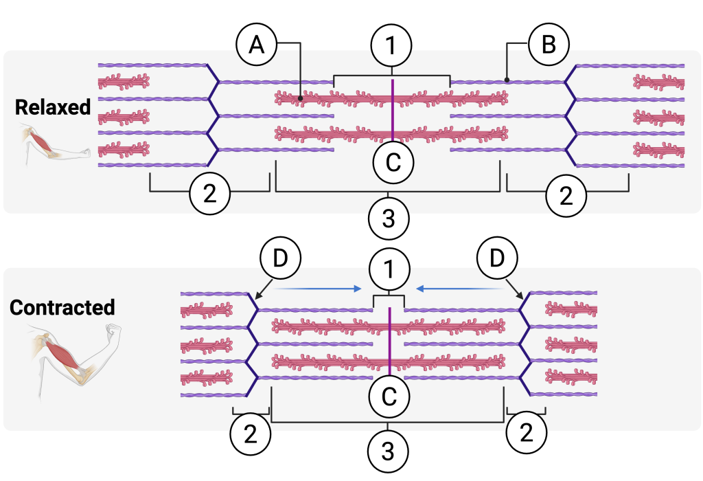
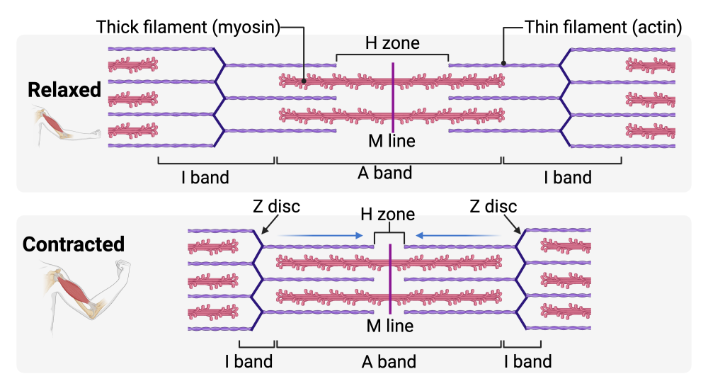
A relaxed sarcomere (top) and a contracted sarcomere (bottom). Click to see a labeled version. - Sarcomere shortening occurs when nerve impulses stimulate muscle cells to release their stores of calcium ions (Ca++). This enables the heads of myosin molecules to attach to binding sites on actin. When the heads pull back — it’s called a power stroke — the sarcomere shortens as the Z lines draw together.
How calcium powers muscle contraction on a molecular level .
Understanding how sarcomere shortening is powered by ATP (and its counterpart, ADP and phosphate) will be the main topic of this tutorial But first, let’s review excitation-contraction coupling.
2. Excitation-Contraction Coupling: A Review
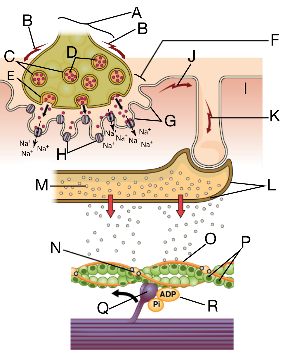
The diagram on the right shows how a nerve impulse gets transformed into a muscle contraction.
- Letter “A” represents the axonal terminal. At “B,” we see the nerve impulse — an action potential — traveling along the membrane of the axon as a wave of membrane depolarization. That action potential induces synaptic vesicles (“C”) filled with the neurotransmitter acetylcholine (“D”), to fuse with the axon’s cell membrane. Through exocytosis (“E”), the acetylcholine is released into the synaptic cleft (“F”), the fluid-filled space in between the axonal bulb and the motor end-plate (“G”): the specialized, folded membrane of the sarcolemma that lies opposite the axonal bulb.
- The acetylcholine diffuses across the synaptic cleft and binds with acetylcholine receptors (“H”) embedded in the motor end plate. These receptors are a part of gated membrane channels that open up in response to the binding of acetylcholine, allowing sodium ions — Na+ — to diffuse from the extracellular fluid into the sarcoplasm (“I”): the cytoplasm of the muscle fiber.
- If enough Na+ diffuses into the muscle fiber, a second action potential — this one happening in the muscle fiber— will start to move along the sarcolemma: this action potential is represented by “J”. This action potential will also move down into the T-tubules (“K”), indentations of the membrane that penetrate deeply into the sarcoplasm.
- In the sarcoplasm, the T-tubules connect to the sarcoplasmic reticulum (“L”).In the sarcoplasmic reticulum (SR), the action potential causes calcium ions (Ca2+, represented by “M”) to diffuse out of the SR and come into contact with the sarcomeres. The Ca2+ will bind with troponin (“N”), a protein that can adjust the position of tropomyosin (“O”), a long, fibrous protein.
- In relaxed muscle tissue, tropomyosin covers the actin-binding sites (“P”) on the thin filaments. But in response to Ca2+, the tropomyosin is nudged aside, The allows the myosin heads (“Q”) to bind with the myosin binding sites on actin.
Below, we’ll see how interactions between the myosin head and ADP and Phosphate (and ATP) generate the force that leads to sarcomere shortening. But first, a quiz.
3. Excitation-Contraction Coupling Quiz
[qwiz use_dataset=”Anatomy Diagrams one letter answers|unit:11.Muscle Tissue|topic:11.4.Excitation-Contraction Coupling” random=”true” style=”width: 600px !important; min-height: 600px !important;” qrecord_id=”sciencemusicvideosMeister1961-Excitation-Contraction Coupling Diagram Quiz”]
[h]Excitation-Contraction Coupling Diagram Quiz
[i]
[x]
[restart]
[/qwiz]
4. ATP, ADP, and the Cross-Bridge Cycle
Moving muscles during exercise is mechanical work. Work requires energy, and in cells, the molecule that powers work is ATP (adenosine triphosphate).
In relation to muscle contraction, here’s what you need to know about ATP.
- ATP is the cell’s main energy-carrying molecule.
- ATP and ADP are two forms of the same molecule, constantly being recycled in cells.
- When ATP loses a phosphate and becomes ADP, energy is released that cells can use to do work. Above, the phosphate is shown by a circled letter “P”. In the discussion below, we’ll represent phosphate as Pi. The subscript “i” stands for inorganic (because the phosphate isn’t connected to any carbon atoms).
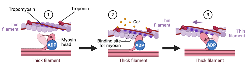
Here’s how ATP and ADP power muscle contraction. The diagram below shows a single myosin head interacting with an actin filament. In a real sarcomere, however, each thick filament contains hundreds of myosin heads, and each head can bind to actin. In addition, each sarcomere contains thousands of overlapping actin and myosin filaments, and each muscle fiber contains thousands of sarcomeres arranged end to end within each myofibril. When all of these interactions occur together, their combined effect is the large force produced by a contracting muscle.
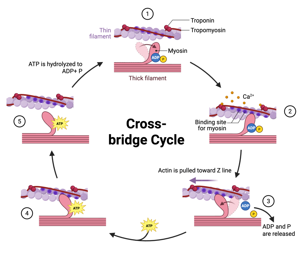
- In diagram 1, we see a representation of myosin and actin in a relaxed sarcomere. The myosin head, which is bound to ADP and inorganic phosphate (Pi), is cocked and ready for action. However, it cannot bind to the thin filament because tropomyosin is blocking the actin-binding sites on actin.
- In diagram 2, calcium ions (Ca2+) appear on the scene. These calcium ions were released from the sarcoplasmic reticulum into the sarcoplasm. The Ca2+ binds to troponin, which causes tropomyosin to shift aside. This exposes the actin-binding sites on the thin filament, allowing the myosin head to form a temporary bond with actin. This bond is called a cross-bridge.
- Diagram 3 shows the power stroke. The myosin head releases inorganic phosphate (Pi), followed by ADP. This triggers a change in the shape of the myosin head, causing it to pivot and pull the actin filament toward the Z line. The combined action of millions of myosin heads pulling on actin filaments is the molecular basis of the force generated by muscle contraction.
- Diagram 4 shows ATP binding to the myosin head.
- Diagram 5 shows the effect of ATP binding: the myosin head detaches from the actin-binding site on the thin filament. The myosin head then hydrolyzes ATP to ADP and Pi using its ATPase activity (an ATPase is an enzyme that can break down ATP). This hydrolysis re-cocks the myosin head into its high-energy position, returning the system to the state shown in diagram 1.
If calcium continues to be released from the sarcoplasmic reticulum, this cycle will repeat, leading the muscle fiber to contract further. The process ends when:
- The nerve impulse ends and calcium ions are actively pumped back into the sarcoplasmic reticulum.
- ATP becomes unavailable (as in extreme fatigue or death, which leads to rigor mortis).
5. Quiz: The Cross Bridge Cycle
[qwiz use_dataset=”Anatomy Diagrams one letter answers|unit:11.Muscle Tissue|topic:11.5.Cross-Bridge Cycle” random=”true” style=”width: 600px !important; min-height: 600px !important;” qrecord_id=”sciencemusicvideosMeister1961-Cross-Bridge Cycle Diagram Quiz”]
[h]Excitation-Contraction Coupling Diagram Quiz
[i]
[x]
[restart]
[/qwiz]
This tutorial ends this module on muscle contraction.