1. Introduction: How can signals travel the length of a neuron?
 In the last tutorial, we looked at reflex arcs, simple systems of connected neurons that enable animals to quickly respond to a stimulus. While you should, at this point, have an understanding of the structure of a reflex arc, we still haven’t addressed the underlying workings. How does this system transmit information?
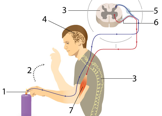
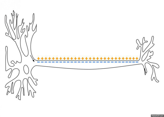
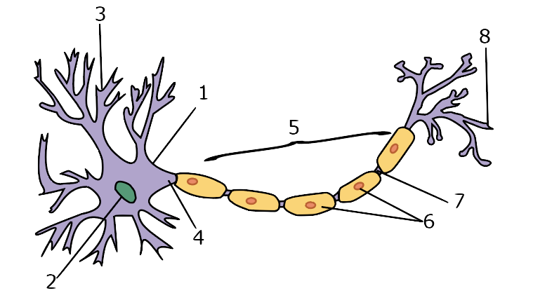
To get a sense of what we need to explain, take another look at the diagram of a reflex arc to your left. When you burn your finger, information has to be conveyed from the tip of your finger (1), all the way to your spinal cord (3). That information has to travel the length of a sensory neuron (5). Then that information (“Burn!”) has to be transmitted across a synapse to a motor neuron (6), which has to send a message (“contract”) to a muscle cell (at 7). So our key question (for now) is this: How can information travel along the length of a neuron?
2. Nerve Impulses: The Big Picture
Let’s start by jumping right to the answer. Click “play” and watch a few seconds from my video “Membranes!”
What this animation is trying to depict is this: a nerve impulse is a wave of ions, diffusing across the membrane of a nerve cell, moving down the length of the nerve cell.
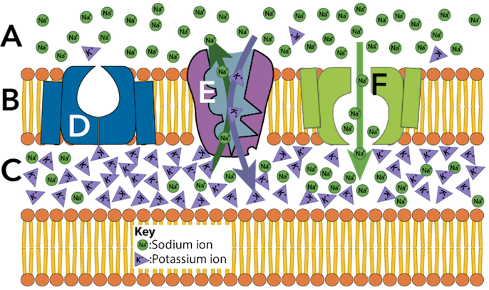
Here’s a diagram that’s representing the same thing. Note that A, B, and C are a time series: they represent the same section of an axon over a few milliseconds (one millisecond = 1/1000 of a second).
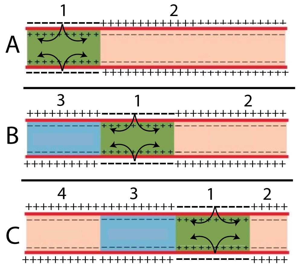 A nerve impulse is like a chain of dominoes: as the dominoes fall in one area (area 1, shown in green in the diagram to the left), they cause the dominoes in the adjacent region (area 2, in tan) to fall. In a neuron, as the positive ions in one region flow in, they cause changes in the membrane that allow the positive ions in the adjacent region of the cell to flow in. The signal that’s traveling from your finger to your spinal cord is this wave of diffusing ions flowing across a sensory neuron’s membrane.
3. Resting potential sets up the conditions for impulses to travel
 Like all cells, neurons maintain an electrical charge across their membrane. To understand what this means, let’s connect this to something you use all the time: an electrical battery. Here are two well known facts about batteries.
- Batteries store electrical energy. That energy is measured as a type of potential energy that’s called voltage.
- Batteries have a positive pole and a negative pole. In the battery shown to the right the positive pole is on top, represented by the plus sign (“+”), while the negative pole is on the bottom, represented by the minus sign (“-“).
So, what is voltage? It’s the potential energy difference between the positive pole and the negative pole. That difference allows you to use the battery to power all kinds of work (such as powering whatever device you’re using). When the voltage disappears, which occurs when a battery goes dead, the battery can no longer power work.
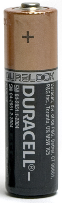
The charge in the AA battery shown above is three volts. You can measure that with a device that’s called a voltmeter (which is, essentially, what a battery tester is).
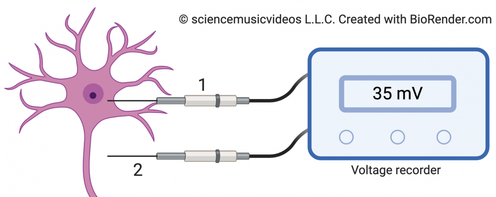
A modified version of a voltmeter can be used to measure the charge across a cell membrane. Here’s our set up:
The voltage recorder has two electrodes. The first (1) gets placed inside the cell’s cytoplasm. The second (2) is the reference electrode: it’s placed outside the cell. In a neuron that’s at rest (which means that it’s not firing an impulse), the voltage recorder will display a voltage of -70 mV (millivolts). The charge is negative because, as we’ll see, a neuron at rest has a negative charge inside the membrane (in the cytoplasm), relative to the outside (the extracellular fluid).
While the difference in charge is measured in voltage, most of what you’ll read below talks about potential, which is short for potential energy. So when you see terms like membrane potential or resting potential, remember that what we’re talking about is voltage: stored electrical energy.
[qwiz qrecord_id=”sciencemusicvideosMeister1961-Resting Potential IR, M28″]
[h]Resting Potential: Interactive Reading
[i]So, why is there a – 70 mV charge across the membrane? The immediate answer is that it’s because the concentrations of various ions are different across the membrane.
Click “continue” to answer the questions that follow.
[q]Let’s start by making sure we can read this diagram correctly. What’s the concentration of sodium (Na+) outside the membrane?
[c]MTUgbU0=[Qq]
[f]Tm8uIDE1IG1NIGlzIHRoZSBjb25jZW50cmF0aW9uIG9mIHNvZGl1bSAocmVwcmVzZW50ZWQgYnkgW05hKw==XSkgaW5zaWRlIHRoZSBtZW1icmFuZS4gRmluZCB0aGUgbnVtYmVyIHRoYXQmIzgyMTc7cyB1bmRlcm5lYXRowqBbTmE=Kw==XSBpbiB0aGUgZXh0cmFjZWxsdWxhciBmbHVpZC4=[Qq]
[c]MTUw IG1N[Qq]
[f]RXhjZWxsZW50LiBUaGUgY29uY2VudHJhdGlvbiBvZiBzb2RpdW0gKHJlcHJlc2VudGVkIGJ5IFtOYQ==Kw==XSkgb3V0c2lkZSB0aGUgbWVtYnJhbmUgaXMgMTUwIG1NLg==[Qq]
[c]MTIwIG1N[Qq]
[f]Tm8uIDEyMCBtTSBpcyB0aGUgY29uY2VudHJhdGlvbiBvZiBwb3Rhc3NpdW0gaW9ucyAocmVwcmVzZW50ZWQgYnkgW0NsJiM4MjExOw==XSkgb3V0c2lkZSB0aGUgbWVtYnJhbmUuIEZpbmQgdGhlIG51bWJlciB0aGF0JiM4MjE3O3MgdW5kZXJuZWF0aMKgW05hKw==XSBpbiB0aGUgZXh0cmFjZWxsdWxhciBmbHVpZC4=[Qq]
[c]NSBtTQ==[Qq]
[f]Tm8uIDUgbU0gaXMgdGhlIGNvbmNlbnRyYXRpb24gb2YgY2hsb3JpbmUgaW9ucyAocmVwcmVzZW50ZWQgYnlbSw==Kw==XSkgb3V0c2lkZSB0aGUgbWVtYnJhbmUuIEZpbmQgdGhlIG51bWJlciB0aGF0JiM4MjE3O3MgdW5kZXJuZWF0aMKgW05hKw==XSBpbiB0aGUgZXh0cmFjZWxsdWxhciBmbHVpZC4=[Qq]
[q]Where are sodium ions (Na+) in higher concentration?
[c]SW5zaWRlIHRoZSBtZW1icmFuZQ==[Qq]
[c]T3V0c2lkZSB0aG UgbWVtYnJhbmU=[Qq]
[f]Tm8uIFRoZSBjb25jZW50cmF0aW9uIGlzIGxpc3RlZCB1bmRlcm5lYXRoIHRoZSBpb24gKGFuZCBhbHNvIGluZGljYXRlZCBieSB0aGUgc2l6ZSBvZiB0aGUgdHlwZWZhY2UpLiBXaGVyZSBhcmUgc29kaXVtIGlvbnMgKE5hKw==KSBtb3JlIGNvbmNlbnRyYXRlZD8=[Qq]
[f]WWVzLiBOYQ==Kw==IGlzIG1vcmUgY29uY2VudHJhdGVkIG91dHNpZGUgdGhlIG1lbWJyYW5lLg==[Qq]
[q]Where are potassium ions (K+) in higher concentration?
[c]SW5zaWRlIHRoZS BtZW1icmFuZQ==[Qq]
[c]T3V0c2lkZSB0aGUgbWVtYnJhbmU=[Qq]
[f]WWVzLiBLKw==IGlzIG1vcmUgY29uY2VudHJhdGVkIGluc2lkZSB0aGUgbWVtYnJhbmUu[Qq]
[f]Tm8uIFRoZSBjb25jZW50cmF0aW9uIGlzIGxpc3RlZCB1bmRlcm5lYXRoIHRoZSBpb24gKGFuZCBhbHNvIGluZGljYXRlZCBieSB0aGUgc2l6ZSBvZiB0aGUgdHlwZWZhY2UpLiBXaGVyZSBpcyBLKw==IG1vcmUgY29uY2VudHJhdGVkPw==[Qq]
[q labels = “top”]
In terms of understanding signaling processes in neurons, the main thing to remember is the relative concentration of sodium and potassium ions. Specifically, you need to remember that ______ ions are more concentrated outside the membrane, and _____ ions are more concentrated inside the membrane. But, in addition, you should notice that there’s a lot more anions (negative ions, represented by ______) in the cytoplasm, and a lot more chlorine, represented by ______ in the extracellular fluid.
[l]A–
[fx] No, that’s not correct. Please try again.
[f*] Correct!
[l]Cl–
[fx] No, that’s not correct. Please try again.
[f*] Correct!
[l]K+
[fx] No. Please try again.
[f*] Excellent!
[l]Na+
[fx] No, that’s not correct. Please try again.
[f*] Correct!
[q]
Note that the 120mM concentration of Chlorine ions (just to pick one example) doesn’t translate to 120 mV. While it’s possible to calculate voltage difference across membranes from ion potential, that’s something that’s outside the scope of a typical AP Biology course, so don’t worry about it. Your job is to remember that the neuron membrane, like a battery, stores ________ energy.
[hangman]
[c]ZWxlY3RyaWNhbA==[Qq]
[/qwiz]
So far, we’ve established that
- Membranes store electrical potential, also known as voltage.
- That potential is related to the difference in ion concentrations across the membrane.
- In a neuron that’s at rest (which means that it’s not firing a signal), the charge across the membrane is about -70 mV.
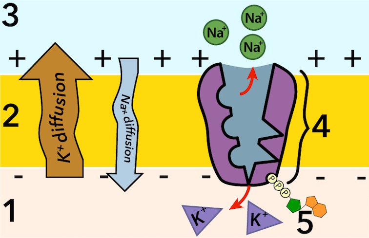
Now let’s dig deeper and ask why these ions are found where they’re found. Note that these ion concentrations represent a profound disequilibrium. When an organism dies, these concentration gradients go away, and everything diffuses to a low energy state. In other words, it takes work to create these concentration gradients.
The source of the energy to power that work is shown at 5 in the diagram to your left. That’s ATP. ATP powers the sodium-potassium pump (4), which pumps sodium ions out of the cytoplasm (1) and into the extracellular fluid (3), while pumping potassium ions from the extracellular fluid and into the cytoplasm. The ratio of this pumping is 3 Na+ pumped out of the cell for every 2 K+ pumped into the cell.
Because of membrane channels that allow sodium ions to diffuse back into the cell and potassium ions to diffuse back out, the sodium-potassium pump is constantly at work. In fact, a third of your ATP (source: protein databank) is used to power this pump, establishing the gradients that make nerve impulses possible.
The membrane channels I just referred to play a key role in establishing the the -70mV resting potential. But for now, we’re going to keep things simple. Just remember that
- The sodium potassium pump is pumping sodium ions out of the cell and potassium ions into the cell. Like all pumping, this requires energy. It’s an endergonic process, powered by hydrolysis of ATP into ADP and Pi (inorganic phosphate).
- Because they’re charged particles, these sodium and potassium ions can’t diffuse through the phospholipid bilayer. There are, however, membrane channels that facilitate the diffusion of each ion across the membrane.
- There are lots of sodium ions just outside the cell membrane that “want” to follow their concentration gradient and flow into the cytoplasm, and lots of potassium ions inside the cytoplasm that “want” to follow their concentration gradient and flow across the membrane and into the extracellular fluid.
That’s it. To make sure you understand, take the following quiz.
[qwiz random = “true” qrecord_id=”sciencemusicvideosMeister1961-Resting Potential, Na_K Pump Quiz, M28″]
[h]Resting Potential and the Sodium-Potassium Pump: Checking Understanding
[i]
[q]Which number in the diagram below represents the cytoplasm?
[textentry single_char=”true”]
[c]MQ ==[Qq]
[f]WWVzLiDigJwx4oCdIGlzIHRoZSBjeXRvcGxhc20u[Qq]
[c]Kg==[Qq]
[f]Tm8uIEhlcmUmIzgyMTc7cyBhIGhpbnQuIFRoZSBzb2RpdW0gcG90YXNzaXVtIHB1bXAgcHVtcHMgTmE=Kw==IGlvbnMgb3V0IG9mIHRoZSBjeXRvcGxhc20sIGFuZCBpbnRvIHRoZSBleHRyYWNlbGx1bGFyIHNwYWNlLg==
Cg==[Qq]
[q]Which number in the diagram below represents the cell membrane of an neuron?
[textentry single_char=”true”]
[c]Mg ==[Qq]
[f]WWVzLiDigJwy4oCdIGlzIHRoZSBjZWxsIG1lbWJyYW5lLg==[Qq]
[c]Kg==[Qq]
[f]Tm8uIEhlcmUmIzgyMTc7cyBhIGhpbnQuIElvbnMgZ2V0IHB1bXBlZCAob3IgZGlmZnVzZWQpIGFjcm9zcyB0aGUgbWVtYnJhbmUu
Cg==[Qq]
[q]Which number in the diagram below represents the extracellular space?
[textentry single_char=”true”]
[c]Mw ==[Qq]
[f]WWVzLiDigJwz4oCdIGlzIHRoZSBleHRyYWNlbGx1bGFyIHNwYWNlLg==[Qq]
[c]Kg==[Qq]
[f]Tm8uIEhlcmUmIzgyMTc7cyBhIGhpbnQuIFRoZSBzb2RpdW0gcG90YXNzaXVtIHB1bXAgKDQpIHB1bXBzIE5hKw==IGlvbnMgb3V0IG9mIHRoZSBjeXRvcGxhc20sIGFuZCBpbnRvIHRoZSBleHRyYWNlbGx1bGFyIHNwYWNlLg==
Cg==[Qq]
[q]Which number in the diagram below represents the sodium potassium pump?
[textentry single_char=”true”]
[c]NA ==[Qq]
[f]WWVzLiDigJw04oCdIGlzIHRoZSBzb2RpdW0gcG90YXNzaXVtIHB1bXAu[Qq]
[c]Kg==[Qq]
[f]Tm8uIEhlcmUmIzgyMTc7cyBhIGhpbnQuIFRoZSBzb2RpdW0gcG90YXNzaXVtIHB1bXAgcHVtcHMgTmE=Kw==IGlvbnMgb3V0IG9mIHRoZSBjZWxsLCBhbmQgSw==Kw==IGlvbnMgaW50byB0aGUgY2VsbC4=
[Qq][q]Which number in the diagram below represents ATP?
[textentry single_char=”true”]
[c]NQ ==[Qq]
[f]WWVzLiDigJw14oCdIGlzIEFUUC4=[Qq]
[c]Kg==[Qq]
[f]Tm8uIEhlcmUmIzgyMTc7cyBhIGhpbnQuIEFUUCBwb3dlcnMgdGhlIHNvZGl1bSBwb3Rhc3NpdW0gcHVtcCAoc2hvd24gYXQgNCku[Qq]
[q]Which number in the diagram represents the region where sodium ions are most highly concentrated?
[textentry single_char=”true”]
[c]Mw ==[Qq]
[f]WWVzLiDigJwz4oCdIGlzIHRoZSBleHRyYWNlbGx1bGFyIGZsdWlkLCBhbmQgdGhhdCYjODIxNztzIHdoZXJlIGEgcmVzdGluZyBuZXVyb24gaGFzIGl0cyBzb2RpdW0gaW9ucyBpbiBoaWdoZXN0IGNvbmNlbnRyYXRpb24u[Qq]
[c]Kg==[Qq]
[f]Tm8uIEhlcmUmIzgyMTc7cyBhIGhpbnQuIFdoZXJlIGRvIHlvdSBzZWUgbW9yZSBzb2RpdW0gaW9uczogaW5zaWRlIG9yIG91dHNpZGUgdGhlIGNlbGw/[Qq]
[q]In the image below, which number represents sodium ions?
[textentry single_char=”true”]
[c]NA ==[Qq]
[f]WWVzLiDigJw04oCdIGlzIGEgc29kaXVtIGlvbi4=[Qq]
[c]Kg==[Qq]
[f]Tm8uIEhlcmUmIzgyMTc7cyBhIGhpbnQuIFRoZSBzb2RpdW0gcG90YXNzaXVtIHB1bXAgcHVtcHMgdGhyZWUgc29kaXVtIGlvbnMgb3V0IG9mIHRoZSBjZWxsIGZvciBldmVyeSB0d28gcG90YXNzaXVtIGlvbnMgdGhhdCBpdCBwdW1wcyBpbnRvIHRoZSBjZWxsLiBXaGVyZSBkbyB5b3Ugc2VlIHRocmVlIHRoaW5ncyBnZXR0aW5nIHB1bXAgdG8/[Qq]
[q]In the image below, which number represents the sodium ion concentration gradient?
[textentry single_char=”true”]
[c]OQ ==[Qq]
[f]WWVzLiDigJw54oCdIGlzIHRoZSBzb2RpdW0gaW9uIGNvbmNlbnRyYXRpb24gZ3JhZGllbnQu[Qq]
[c]Kg==[Qq]
[f]Tm8uIEhlcmUmIzgyMTc7cyBhIGhpbnQuIEluIGEgcmVzdGluZyBuZXVyb24sIHNvZGl1bSBpb25zIGFyZSBtb3JlIGNvbmNlbnRyYXRlZCBvdXRzaWRlIHRoZSBjZWxsIHRoYW4gaW5zaWRlLiBXaGljaCBudW1iZXIgcmVwcmVzZW50cyB0aGF0Pw==[Qq]
[q]In the image below, which number represents the molecule that’s powering the sodium potassium pump?
[textentry single_char=”true”]
[c]Ng ==[Qq]
[f]WWVzLiDigJw24oCdIGlzIEFUUCwgd2hpY2ggcG93ZXJzIHRoZSBzb2RpdW0gcG90YXNzaXVtIHB1bXAu[Qq]
[c]Kg==[Qq]
[f]Tm8uIEhlcmUmIzgyMTc7cyBhIGhpbnQuIFRoaXMgc2FtZSBtb2xlY3VsZSBwb3dlcnMgbW9zdCBlbmRlcmdvbmljIHByb2Nlc3NlcyBpbiBjZWxscy4gSXQgY29uc2lzdHMgb2YgYSBzdWdhciBjb25uZWN0ZWQgdG8gdGhyZWUgcGhvc3BoYXRlIGdyb3VwcyAoYW5kIGEgbml0cm9nZW5vdXMgYmFzZSwgd2hpY2ggaXMgbm90IHNob3duKS4gV2hhdCBtb2xlY3VsZSBhYm92ZSBjb3VsZCBmaXQgdGhhdCBkZXNjcmlwdGlvbj8=[Qq]
[x][restart]
[/qwiz]
4. Action Potential: Overview
The sodium-potassium pump, and the resting potential that it creates, is a common feature of cells. What makes neurons unique is
- The long processes (dendrites and axons) that extend from the cell body
- Their ability to quickly reverse the polarity of a stretch of membrane, and then have that switched polarity propagate itself along the length of the nerve cell.
This switch in polarity is called an action potential, and we’ll spend the rest of this tutorial seeing how it works.
5. Gated Ion Channels
Along the length of their membranes, neurons have gated ion channels. There are three things to know about them:
- As their name indicates, these channels have gates that can open up in response to certain conditions.
- These channels allow for facilitated diffusion. The previously established concentration gradients (set up by the sodium-potassium pump) are what power the movement of ions through these channels.
- The channels are (for the most part) specific. Some gates will let only sodium ions pass through. Others will let only potassium ions through.
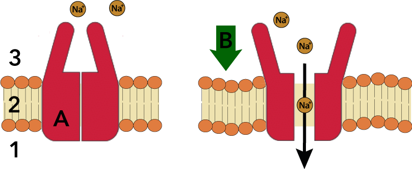 There are only a few types of channels to know about. The channel shown at right is a pressure gated channel (A). Its default condition is closed (as shown on the left side of the diagram). As with any gate, “closed” means that things can’t pass through. In this case, what can’t pass through are ions.
When the membrane (2) is subject to pressure (the green arrow at B), the channel changes shape, allowing ions to flow across the membrane. Remember that unlike the sodium-potassium pump, the ions aren’t being pumped: they’re just following their concentration gradient.
The pressure gated channel shown above is an example of a variety of channels that open in response to touch, heat, cold, or tissue damage. The basic idea is that in response to some stimulus, these channels open, and ions can diffuse through.
 A second type of gated ion channel is a ligand gated ion channel (represented by D, at left). These channels have receptors (E) that bind with a ligand (C). As we’ll see, the ligands involved in the nervous system are neurotransmitters. Binding causes the channel to change its shape in a way that allows a specific ion to flow through.
A third type of gated ion channel is a voltage gated channel.
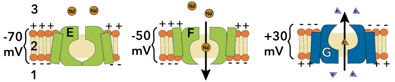 These channels respond to changes in voltage. For example, the voltage gated sodium channel shown at E is a sodium ion channel that’s closed when the membrane is polarized at its resting potential (-70 mV). However, when the polarity decreases to -50mV, the gate opens, allowing sodium ions to diffuse through (as shown at F). Another voltage gated channel is shown at G. This channel opens when the polarity of the membrane rises to +30 mV. At that voltage, the channel opens to potassium ions.
6. Hyperpolarizations and Depolarizations
Because of these gated ion channels, segments of neuronal membranes can be hyperpolarized or depolarized.
- In a hyperpolarization, the membrane becomes more polarized than it is during the resting potential (-70 mV). That means that the charge across the membrane would become more negative (-75mV, -80 mV, etc.)
- In a depolarization, the membrane becomes less polarized than it is during the resting potential. That means that the charge across the membrane would become less negative (-60 mV, -30 mV, 10 mV, 30 mV, etc.)
It’s important that you understand this, so let’s interact with these ideas a bit.
[qwiz random = “true” qrecord_id=”sciencemusicvideosMeister1961-Hyper and Depolarizations, M28″]
[h]Hyperpolarizations and depolarizations
[q]The image on the far left shows ion concentrations when the neuron is at its resting potential. Which graph would result if pressure-gated sodium ion channels were to open, allowing sodium ions to diffuse down their concentration gradient?
[c]McKgIMKgIMKgIMKgIA==[Qq][c]Mg ==
Cg==[Qq][f]Tm8uIEdyYXBoIDEgaXMgc2hvd2luZyB0aGUgY2hhcmdlIGFjcm9zcyB0aGUgbWVtYnJhbmUgYmVjb21pbmcgbW9yZSBuZWdhdGl2ZSAoZHJvcHBpbmcgYmVsb3cgdGhlIC03MCBtViB2YWx1ZSB0aGF0IGl0IGhhcyBkdXJpbmcgcmVzdGluZyBwb3RlbnRpYWwpLiBXaGF0IG9jY3VycyB3aGVuIHNvZGl1bSBpb24gY2hhbm5lbHMgb3BlbiBpcyB0aGUgb3Bwb3NpdGUuIEhlcmUmIzgyMTc7cyBob3cgdG8gdGhpbmsgYWJvdXQgaXQuIEF0IHJlc3RpbmcgcG90ZW50aWFsLCBOYQ==Kw==IGlzIG1vcmUgY29uY2VudHJhdGVkIG91dHNpZGUgdGhlIGNlbGwgdGhhbiBpbnNpZGUuIElmIE5hKw==IGNoYW5uZWxzIG9wZW4sIHRoYXQgbWVhbnMgdGhhdCBOYQ==[Qq]+ will flow into the cell. If positive ions flow into the cytoplasm, will that make the charge more negative or more positive?
[f]RXhjZWxsZW50LiBJZsKgTmE=K8KgZmxvd3MgaW50byB0aGUgY2VsbCwgaXQgd2lsbCBicmluZyBpbiBwb3NpdGl2ZSBjaGFyZ2VzLCBhbmQgdGhlcmVmb3JlIG1ha2UgdGhlIG1lbWJyYW5lIHBvdGVudGlhbCBsZXNzIG5lZ2F0aXZlLiBJbiBvdGhlciB3b3JkcywgaXQmIzgyMTc7cyBjYXVzaW5nIGEgcGFydGlhbCBkZXBvbGFyaXphdGlvbi4=[Qq]
[q]The image on the far left shows ion concentrations when the neuron is at its resting potential. Which graph would result if gated potassium ion channels described above were to open, allowing K+ ions to diffuse down their concentration gradient?
[c]McKgIMKg IMKgIMKg[Qq][c]Mg==
Cg==[Qq][f]RmFidWxvdXMuIEsrIGlzIG1vcmUgY29uY2VudHJhdGVkIGluc2lkZSB0aGUgY2VsbC4gSWYgcG90YXNzaXVtIGdhdGVkIGlvbiBjaGFubmVscyBvcGVuLCB0aGUgcG90YXNzaXVtIGlvbnMgd2lsbCBkaWZmdXNlIG91dCBvZiB0aGUgY2VsbCwgY2FycnlpbmcgdGhlaXIgcG9zaXRpdmUgY2hhcmdlIGZyb20gdGhlIGN5dG9wbGFzbSB0byB0aGUgZXh0cmFjZWxsdWxhciBmbHVpZC4gVGhpcyB3aWxsIG1ha2UgdGhlIG1lbWJyYW5lIHBvdGVudGlhbCBtb3JlIG5lZ2F0aXZlLCBzb21ldGhpbmcgdGhhdCBpcyBrbm93biBhcyBhIA==aHlwZXJwb2xhcml6YXRpb24=Lg==[Qq]
[f]Tm8uIElmIEs=Kw==IGNoYW5uZWxzIG9wZW4sIHRoZW4gSw==Kw==IHdpbGwgZmxvdyBvdXQsIGNhcnJ5aW5nIHBvc2l0aXZlIGNoYXJnZSBmcm9tIHRoZSBjeXRvcGxhc20gdG8gdGhlIGV4dHJhY2VsbHVsYXIgZmx1aWQuIElmIHlvdSBsb3NlIHNvbWV0aGluZyBwb3NpdGl2ZSwgeW91IG1ha2UgaXQgbW9yZSBfX19fX19fX18/[Qq]
[q]If K+ gated ion channels open, which of the following readings in the voltage recorder is most likely?
[c]LTcwIG1WwqAgwqAgwqA=[Qq][c]LTUwIG12wqAgwqAgwqA=[Qq][c]LTgw IG12
Cg==[Qq][f]Tm8uIFRoZSByZXN0aW5nIHBvdGVudGlhbCBpcyBhdCAtNzAgbVYuIElmIGEgcG90YXNzaXVtIGdhdGVkIGlvbiBjaGFubmVscyB3ZXJlIHRvIG9wZW4sIHBvdGFzc2l1bSBpb25zIHdvdWxkIGZsb3cgdGhyb3VnaCB0aGVtLCBhbmQgdGhlIG1lbWJyYW5lIHBvdGVudGlhbCB3b3VsZCBjaGFuZ2UuIFdvdWxkIGl0IGdldCBoaWdoZXIgb3IgbG93ZXI/[Qq]
[f]Tm8uIFRoZSByZXN0aW5nIHBvdGVudGlhbCBpcyBhdCAtNzAgbVYuIElmIGEgcG90YXNzaXVtIGdhdGVkIGNoYW5uZWwgd2VyZSB0byBvcGVuLCBwb3Rhc3NpdW0gaW9ucyB3b3VsZCBmbG93IHRocm91Z2ggaXQsIGFuZCBjYXJyeSB0aGVpciBwb3NpdGl2ZSBjaGFyZ2UgZnJvbSBpbnNpZGUgdGhlIG1lbWJyYW5lICh3aGVyZSBwb3Rhc3NpdW0gaXMgbW9yZSBjb25jZW50cmF0ZWQpIHRvIG91dHNpZGUgdGhlIG1lbWJyYW5lLiBJZiB0aGUgY3l0b3BsYXNtIHdlcmUgdG8gbG9zZSB0aGVzZSBwb3NpdGl2ZSBjaGFyZ2VzLCBob3cgd291bGQgdGhhdCBhZmZlY3QgdGhlIGNoYXJnZSBhY3Jvc3MgdGhlIG1lbWJyYW5lPw==[Qq]
[f]R3JlYXQgam9iLiBJZiBhIHBvdGFzc2l1bSBnYXRlZCBjaGFubmVsIHdlcmUgdG8gb3BlbiwgcG90YXNzaXVtIGlvbnMgd291bGQgZmxvdyB0aHJvdWdoIGl0LCBjYXJyeWluZyB0aGVpciBwb3NpdGl2ZSBjaGFyZ2UgZnJvbSBpbnNpZGUgdGhlIG1lbWJyYW5lICh3aGVyZSBwb3Rhc3NpdW0gaXMgbW9yZSBjb25jZW50cmF0ZWQpIHRvIG91dHNpZGUgdGhlIG1lbWJyYW5lLiBUaGF0IHdvdWxkIG1ha2UgdGhlIGN5dG9wbGFzbSBtb3JlIG5lZ2F0aXZlIHJlbGF0aXZlIHRvIHRoZSBleHRyYWNlbGx1bGFyIGZsdWlkLiBUaGUgbWVtYnJhbmUgcG90ZW50aWFsIHdvdWxkIHRodXMgZGVjcmVhc2UgZnJvbSBpdHMgcmVzdGluZyB2YWx1ZSBvZiAtNzAgbVYgYW5kIG1vdmUgdG8gLTgwIG1WLg==[Qq]
[q]If Na+ gated ion channels open, which of the following readings in the voltage recorder is most likely?
[c]LTcwIG1WwqAgwqAgwqA=[Qq][c]LTUwIG12wq AgwqAgwqA=[Qq][c]LTgwIG12
Cg==[Qq][f]Tm8uIFRoZSByZXN0aW5nIHBvdGVudGlhbCBpcyBhdCBhYm91dCAtNzAgbVYuIElmIHNvZGl1bSBnYXRlZCBpb24gY2hhbm5lbHMgd2VyZSB0byBvcGVuLCBzb2RpdW0gaW9ucyB3b3VsZCBmbG93IHRocm91Z2ggaXQsIGFuZCB0aGUgbWVtYnJhbmUgcG90ZW50aWFsIHdvdWxkIGNoYW5nZS4gV291bGQgaXQgZ2V0IGhpZ2hlciBvciBsb3dlcj8=[Qq]
[f]R3JlYXQgam9iLiBUaGUgcmVzdGluZyBwb3RlbnRpYWwgaXMgYXQgYWJvdXQgLTcwIG1WLiBJZiBnYXRlZCBzb2RpdW0gaW9uIGNoYW5uZWxzIHdlcmUgdG8gb3BlbiwgdGhlbiBzb2RpdW0gaW9ucyB3b3VsZCBmbG93IGRvd24gdGhlaXIgY29uY2VudHJhdGlvbiBncmFkaWVudCwgYW5kIGZsb3cgaW50byB0aGUgY2VsbC4gQXMgdGhleSBkaWQsIHRoZXkmIzgyMTc7ZCBicmluZyBwb3NpdGl2ZSBjaGFyZ2UgaW50byB0aGUgY3l0b3BsYXNtLCB3aGljaCB3b3VsZCBkZWNyZWFzZSB0aGUgbWVtYnJhbmUgcG90ZW50aWFsIChtYWtpbmcgaXQgbGVzcyBuZWdhdGl2ZSBhbmQgbW9yZSBwb3NpdGl2ZSku[Qq]
[f]Tm8uIFRoZSByZXN0aW5nIHBvdGVudGlhbCBpcyBhdCBhYm91dCAtNzAgbVYuIElmIGdhdGVkIHNvZGl1bSBpb24gY2hhbm5lbHMgd2VyZSB0byBvcGVuLCB0aGVuIHNvZGl1bSBpb25zIHdvdWxkIGZsb3cgZG93biB0aGVpciBjb25jZW50cmF0aW9uIGdyYWRpZW50LCBhbmQgZmxvdyBpbnRvIHRoZSBjZWxsLiBBcyB0aGV5IGRpZCwgdGhleSYjODIxNztkIGJyaW5nIHBvc2l0aXZlIGNoYXJnZSBpbnRvIHRoZSBjeXRvcGxhc20uIFdvdWxkIHRoYXQgbWFrZSB0aGUgbWVtYnJhbmUgcG90ZW50aWFsIG1vcmUgcG9zaXRpdmUsIG9yIG1vcmUgbmVnYXRpdmU/[Qq]
[q labels = “top”](Image from Campbell Biology, Pearson)
[l]De
[fx] No, that’s not correct. Please try again.
[f*] Great!
[l]Hyper
[fx] No. Please try again.
[f*] Excellent!
[l]K+
[fx] No, that’s not correct. Please try again.
[f*] Excellent!
[l]Na+
[fx] No. Please try again.
[f*] Correct!
[x][restart]
[/qwiz]
7. Action Potential: The Details
The depolarizations and hyperpolarizations that we studied above only changed the polarity of the membrane by a small amount. That amount is proportional to the strength of the stimulus. A stimulus that decreases the membrane potential results in a graded depolarization. A stimulus with the opposite effect (increasing the membrane potential) results in a graded hyperpolarization.
In an action potential, the shift in polarity takes the membrane’s charge from -70 mV to a positive value (about 35 mV). If we were to record the voltage across a segment of membrane during an action potential, here’s what we’d see a few milliseconds into the process.
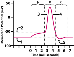 Now, imagine that we were to graph the numbers on the voltage recorder over the few milliseconds that pass during an action potential. The image on the right shows what our graph would look like. X axis is time, and Y axis is membrane potential, in millivolts.
Before reading the description below, ask yourself a few questions.
- Which number is resting potential?
- When are sodium gates opening?
- When are potassium gates opening?
Now, read what’s below.
|
 |
|
In the graph of action potential on the left, number 1 indicates the membrane at resting potential, about -70 mV. At this moment, because of the work of the sodium-potassium pump (E, in the diagram on the right), sodium ions predominate in the extracellular fluid and potassium ions predominate in the cytoplasm.
At 0 milliseconds, a stimulus causes gated ion channels to open. We know that these are sodium channels, because they’re causing a depolarization (the membrane potential is becoming less negative).
As mentioned above, the depolarization that’s happening between 0 and 2 milliseconds, indicated by letter “A,” is a graded depolarization. That means that the change in polarity is proportional to the strength of the stimulus. As the stimulus continues, the polarity continues to decrease, until it reaches the dotted line at “2.” The value, at that point, is about -50 mV.
|
 |
|
-50 mV is the threshold potential. At that voltage, voltage-gated sodium channels (F) open up, allowing for an influx of sodium ions to cross the membrane and enter the cytoplasm. The threshold represents the initiation of what’s called an all-or-none event, which means that once the membrane depolarizes to -50 mV, all of the voltage-gated sodium channels in that patch of membrane open. This abrupt, inward flow of sodium ions is called the rising phase of the the action potential (shown at 3 in the graph, above left). Within a millisecond or two, the membrane potential changes from negative to positive, peaking at about 35 mV.
|
 |
|
That peak (35 mV) acts as a stimulus for two things to happen.
- The voltage sensitive sodium ion gates that caused the depolarization close.
- Voltage sensitive potassium ion gates ion open (D). As these channels open, potassium ions rush out of the cell. As these positive ions leave the cell, the membrane potential falls from 35 mV back into negative territory. That drop in voltage is represented by 4 in the graph and it’s called the falling phase of the action potential.
|
 |
|
Now we’ve covered the graded depolarization (at “A”), which leads to the action potential (at “B”). The action potential ends in a refractory period (“C”). During the refractory period, potassium ions have left the cell to such an extent that the membrane becomes hyperpolarized: its charge is lower than it was during the resting potential, dropping to about -80 mV. For this reason, the refractory period is also called the undershoot. During these few milliseconds, that portion of the membrane can’t undergo another action potential. Part of the reason for that is that the action potential is based on a previously established resting potential, and the resting potential hasn’t, at this point, been re-established. There are still lots of sodium ions inside the cytoplasm, and lots of potassium ions in the extracellular fluid. To reestablish the resting potential, the sodium-potassium pump is going to need a few milliseconds to do its work.
In a moment, we’ll see how this action potential can propagate itself down the length of an axon. But first, let’s consolidate our understanding of what’s above.
[qwiz random = “true” qrecord_id=”sciencemusicvideosMeister1961-Action Potential Quiz, M28″] [h]
Action Potential Quiz
[i]
[q] In the diagram below, which number or letter represents the resting potential?
[textentry single_char=”true”]
[c]ID E=[Qq]
[f]IEV4Y2VsbGVudC4gUmVzdGluZyBwb3RlbnRpYWwgaXMgcmVwcmVzZW50ZWQgYnkgbnVtYmVyIDEu[Qq]
[c]IEVudGVyIHdvcmQ=[Qq]
[f]IFNvcnJ5LCB0aGF0JiM4MjE3O3Mgbm90IGNvcnJlY3Qu[Qq]
[c]ICo=[Qq]
[f]IE5vLiBIZXJlJiM4MjE3O3MgYSBoaW50LiBUaGUgcmVzdGluZyBwb3RlbnRpYWwgcHJlY2VkZXMgYW5kIGZvbGxvd3MgdGhlIGFjdGlvbiBwb3RlbnRpYWwu[Qq]
[q] In the diagram below, which number or letter represents the threshold potential?
[textentry single_char=”true”]
[c]ID I=[Qq]
[f]IE5pY2Ugam9iLiBUaHJlc2hvbGQgcG90ZW50aWFsIGlzIHJlcHJlc2VudGVkIGJ5IG51bWJlciAyLg==[Qq]
[c]IEVudGVyIHdvcmQ=[Qq]
[f]IFNvcnJ5LCB0aGF0JiM4MjE3O3Mgbm90IGNvcnJlY3Qu[Qq]
[c]ICo=[Qq]
[f]IE5vLiBIZXJlJiM4MjE3O3MgYSBoaW50LiBUaGUgdGhyZXNob2xkIHBvdGVudGlhbCBpcyB0aGUgcG90ZW50aWFsIGJleW9uZCB3aGljaCBhbiBhY3Rpb24gcG90ZW50aWFsIHN0YXJ0cy4gU2VlIGlmIHlvdSBjYW4gZmluZCB3aGVyZSB0aGUgYWN0aW9uIHBvdGVudGlhbCBzdGFydHMsIGFuZCB0aGVuIGZpbmQgdGhlIGNvcnJlc3BvbmRpbmcgbGluZSBvbiB0aGUgWSBheGlzLg==[Qq]
[q] In the diagram below, which number or letter represents the rising phase of the action potential?
[textentry single_char=”true”]
[c]ID M=[Qq]
[f]IFRlcnJpZmljLiBUaGUgcmlzaW5nIHBoYXNlIG9mIHRoZSBhY3Rpb24gcG90ZW50aWFsIGlzIHJlcHJlc2VudGVkIGJ5IG51bWJlciAzLg==[Qq]
[c]IEVudGVyIHdvcmQ=[Qq]
[f]IFNvcnJ5LCB0aGF0JiM4MjE3O3Mgbm90IGNvcnJlY3Qu[Qq]
[c]ICo=[Qq]
[f]IE5vLiBIZXJlJiM4MjE3O3MgYSBoaW50LiBUaGUgcmlzaW5nIHBoYXNlIG9mIHRoZSBhY3Rpb24gcG90ZW50aWFsIGlzIHdoZW4gc29kaXVtIGlvbnMgYXJlIGRpZmZ1c2luZyBhY3Jvc3MgdGhlIG1lbWJyYW5lLCBjYXVzaW5nIHRoZSBtZW1icmFuZSYjODIxNztzIHBvdGVudGlhbCB0byByYXBpZGx5IHNoaWZ0IGZyb20gbmVnYXRpdmUgdG8gcG9zaXRpdmUu[Qq]
[q] In the diagram below, which number or letter represents the moment when all of the voltage gated sodium ion channels are open, and sodium is freely diffusing across the membrane into the cytoplasm?
[textentry single_char=”true”]
[c]ID M=[Qq]
[f]IFRlcnJpZmljLiBBdCAzLCBkdXJpbmcgdGhlIHJpc2luZyBwaGFzZSBvZiB0aGUgYWN0aW9uIHBvdGVudGlhbCwgYWxsIHZvbHRhZ2UgZ2F0ZWQgc29kaXVtIGNoYW5uZWxzIHdvdWxkIGJlIG9wZW4u[Qq]
[c]IEVudGVyIHdvcmQ=[Qq]
[f]IFNvcnJ5LCB0aGF0JiM4MjE3O3Mgbm90IGNvcnJlY3Qu[Qq]
[c]ICo=[Qq]
[f]IE5vLiBIZXJlJiM4MjE3O3MgYSBoaW50LiBXaGVuIHRoZSB2b2x0YWdlIGdhdGVkIHNvZGl1bSBjaGFubmVscyBhcmUgb3BlbiwgeW91JiM4MjE3O2xsIHNlZSB0aGUgbWVtYnJhbmUmIzgyMTc7cyBwb3RlbnRpYWwgc3RlZXBseSByaXNlIGZyb20gbmVnYXRpdmUgdG8gcG9zaXRpdmUsIGFzIHRoZXNlIHBvc2l0aXZlIGlvbnMgcG91ciBpbnRvIHRoZSBjeXRvcGxhc20u[Qq]
[q] In the diagram below, which number or letter represents the falling phase of the action potential?
[textentry single_char=”true”]
[c]ID Q=[Qq]
[f]IEF3ZXNvbWUuIE51bWJlciA0IHJlcHJlc2VudHMgdGhlIGZhbGxpbmcgcGhhc2Ugb2YgdGhlIGFjdGlvbiBwb3RlbnRpYWwu[Qq]
[c]IEVudGVyIHdvcmQ=[Qq]
[f]IFNvcnJ5LCB0aGF0JiM4MjE3O3Mgbm90IGNvcnJlY3Qu[Qq]
[c]ICo=[Qq]
[f]IE5vLiBIZXJlJiM4MjE3O3MgYSBoaW50LiBUaGUgYWN0aW9uIHBvdGVudGlhbCBpcyB0aGUgbW9tZW50IG9mIHRoZSBiaWdnZXN0IHN3aW5ncyBpbiBtZW1icmFuZSBwb3RlbnRpYWwuIFdoZW4sIGR1cmluZyB0aGUgYWN0aW9uIHBvdGVudGlhbCwgaXMgdGhlIG1lbWJyYW5lIHBvdGVudGlhbCByYXBpZGx5IGRlY3JlYXNpbmc/[Qq]
[q] In the diagram below, which number or letter represents the earliest moment when voltage-gated potassium ion channels are open, allowing potassium to rush out of the cell?
[textentry single_char=”true”]
[c]ID Q=[Qq]
[f]IEV4Y2VsbGVudC4gTnVtYmVyIDQgcmVwcmVzZW50cyB0aGUgZmFsbGluZyBwaGFzZSBvZiB0aGUgYWN0aW9uIHBvdGVudGlhbCwgYW5kIHRoYXQmIzgyMTc7cyB3aGVuwqB2b2x0YWdlLWdhdGVkIHBvdGFzc2l1bSBpb24gY2hhbm5lbHMgYXJlIG9wZW4sIGFsbG93aW5nIHBvdGFzc2l1bSB0byBydXNoIG91dCBvZiB0aGUgY2VsbC4=[Qq]
[c]IEVudGVyIHdvcmQ=[Qq]
[f]IFNvcnJ5LCB0aGF0JiM4MjE3O3Mgbm90IGNvcnJlY3Qu[Qq]
[c]ICo=[Qq]
[f]IE5vLiBIZXJlJiM4MjE3O3MgYSBoaW50LiBXaGVuIHBvdGFzc2l1bSBpb25zIHJ1c2ggb3V0IG9mIHRoZSBjZWxsLCB0aGV5JiM4MjE3O3JlIGNhcnJ5aW5nIHBvc2l0aXZlIGNoYXJnZS4gSWYgcG9zaXRpdmUgY2hhcmdlIGlzIGxlYXZpbmcsIHdpbGwgdGhlIG1lbWJyYW5lIHBvdGVudGlhbCBiZWNvbWUgbW9yZSBwb3NpdGl2ZSBvciBtb3JlIG5lZ2F0aXZlPw==[Qq]
[q] In the diagram below, which number represents the undershoot?
[textentry single_char=”true”]
[c]ID U=[Qq]
[f]IEV4Y2VsbGVudC4gTnVtYmVyIDUgcmVwcmVzZW50cyB0aGUgdW5kZXJzaG9vdCAodGhlIG1vbWVudCB3aGVuIHRoZSBtZW1icmFuZSBwb3RlbnRpYWwgaGFzIGZhbGxlbiBiZWxvdyB0aGUgbGV2ZWwgdGhhdCBpdCYjODIxNztzIGF0IGR1cmluZyByZXN0aW5nIHBvdGVudGlhbCku[Qq]
[c]IEVudGVyIHdvcmQ=[Qq]
[f]IFNvcnJ5LCB0aGF0JiM4MjE3O3Mgbm90IGNvcnJlY3Qu[Qq]
[c]ICo=[Qq]
[f]IE5vLiBIZXJlJiM4MjE3O3MgYSBoaW50LiBUaGUgdW5kZXJzaG9vdCBpcyB0aGUgbW9tZW50IHdoZW4gdGhlIG1lbWJyYW5lIHBvdGVudGlhbCBoYXMgZmFsbGVuIGJlbG93IHRoZSBsZXZlbCB0aGF0IGl0JiM4MjE3O3MgYXQgZHVyaW5nIHJlc3RpbmcgcG90ZW50aWFsLg==[Qq]
[q] In the diagram below, which number or letter represents a graded depolarization?
[textentry single_char=”true”]
[c]IE E=[Qq]
[f]IE5pY2Ugam9iISBMZXR0ZXIgQSByZXByZXNlbnRzIGEgZ3JhZGVkIGRlcG9sYXJpemF0aW9uLg==[Qq]
[c]IEVudGVyIHdvcmQ=[Qq]
[f]IFNvcnJ5LCB0aGF0JiM4MjE3O3Mgbm90IGNvcnJlY3Qu[Qq]
[c]ICo=[Qq]
[f]IE5vLiBIZXJlJiM4MjE3O3MgYSBoaW50LiBUaGUgZ3JhZGVkIGRlcG9sYXJpemF0aW9uIGlzIGEgbW9tZW50IHdoZW4gdGhlIG1lbWJyYW5lIHBvdGVudGlhbCBpcyBtb3ZpbmcgdG93YXJkLCBidXQgaGFzbiYjODIxNzt0IHlldCBleGNlZWRlZCB0aGUgdGhyZXNob2xkIHBvdGVudGlhbC4=[Qq]
[q] In the diagram below, which number or letter represents the action potential?
[textentry single_char=”true”]
[c]IE I=[Qq]
[f]TmljZWx5IERvbmUhIExldHRlciBCIHJlcHJlc2VudHMgdGhlIGFjdGlvbiBwb3RlbnRpYWwu[Qq]
[c]IEVudGVyIHdvcmQ=[Qq]
[f]IFNvcnJ5LCB0aGF0JiM4MjE3O3Mgbm90IGNvcnJlY3Qu[Qq]
[c]ICo=[Qq]
[f]IE5vLiBIZXJlJiM4MjE3O3MgYSBoaW50LiBUaGUgYWN0aW9uIHBvdGVudGlhbCBpcyB3aGVuIHRoZXJlJiM4MjE3O3MgdGhlIG1vc3QgZHJhbWF0aWMgY2hhbmdlIGluIG1lbWJyYW5lIHBvdGVudGlhbC4gV2hlcmUgZG8geW91IHNlZSB0aGF0IGhhcHBlbmluZz8=[Qq]
[q] In the diagram below, which LETTER represents the refractory period?
[textentry single_char=”true”]
[c]Qw ==[Qq]
[f]QXdlc29tZSEgTGV0dGVyIEMgcmVwcmVzZW50cyB0aGUgcmVmcmFjdG9yeSBwZXJpb2Qu[Qq]
[c]IEVudGVyIHdvcmQ=[Qq]
[f]IFNvcnJ5LCB0aGF0JiM4MjE3O3Mgbm90IGNvcnJlY3Qu[Qq]
[c]ICo=[Qq]
[f]IE5vLiBIZXJlJiM4MjE3O3MgYSBoaW50LiBUaGUgcmVmcmFjdG9yeSBwZXJpb2QgaXMgYXNzb2NpYXRlZCB3aXRoIHRoZSB1bmRlcnNob290LiBXaGVyZSBkbyB5b3Ugc2VlIHNvbWV0aGluZyB0aGF0IGxvb2tzIGxpa2UgYW4gdW5kZXJzaG9vdC4=[Qq]
[q]During an intracellular recording of voltage, a student gets the result below.
This reading corresponds with which number on the following graph?
[textentry single_char=”true”]
[c]MQ ==[Qq]
[f]R29vZCB3b3JrISBOdW1iZXIgMSByZXByZXNlbnRzIHRoZSByZXN0aW5nIHBvdGVudGlhbCwgYW5kIHRoYXQmIzgyMTc7cyB3aGVuIHRoZSBtZW1icmFuZSBwb3RlbnRpYWwgaXMgYWJvdXQgLTcwbVYu[Qq]
[c]IEVudGVyIHdvcmQ=[Qq]
[f]IFNvcnJ5LCB0aGF0JiM4MjE3O3Mgbm90IGNvcnJlY3Qu[Qq]
[c]ICo=[Qq]
[f]IE5vLiBMb29rIGF0IHRoZSByZWFkaW5nIG9uIHRoZSB2b2x0bWV0ZXIsIGFuZCB0aGVuIGZpbmQgdGhlIGNvcnJlc3BvbmRpbmcgdmFsdWUgb24gdGhlIGdyYXBoLg==[Qq]
[q]The image below shows a segment of neuronal membrane and a graph of action potential. Which number on the graph best fits the image of the membrane?
[textentry single_char=”true”]
[c]MQ ==[Qq]
[f]R29vZCB3b3JrISBOdW1iZXIgMSByZXByZXNlbnRzIHRoZSByZXN0aW5nIHBvdGVudGlhbC4gU29kaXVtIGlvbnMgYXJlIG1vcmUgY29uY2VudHJhdGVkIG91dHNpZGUgdGhlIG1lbWJyYW5lLCBwb3Rhc3NpdW0gaXMgbW9yZSBjb25jZW50cmF0ZWQgaW5zaWRlIHRoZSBtZW1icmFuZSwgYW5kIGFsbCB2b2x0YWdlIGdhdGVkIGlvbiBjaGFubmVscyBhcmUgY2xvc2VkLg==[Qq]
[c]ICo=[Qq]
[f]IE5vLiBXaGVuIHdvdWxkIHlvdSBoYXZlIGEgc2l0dWF0aW9uIHdoZXJlwqBzb2RpdW0gaW9ucyBhcmUgbW9yZSBjb25jZW50cmF0ZWQgb3V0c2lkZSB0aGUgbWVtYnJhbmUsIHBvdGFzc2l1bSBpcyBtb3JlIGNvbmNlbnRyYXRlZCBpbnNpZGUgdGhlIG1lbWJyYW5lLCBhbmQgYWxsIHZvbHRhZ2UgZ2F0ZWQgaW9uIGNoYW5uZWxzIGFyZSBjbG9zZWQu[Qq]
[q]The image below shows a segment of neuronal membrane and a graph of action potential. Which number on the graph best fits the image of the membrane?
[textentry single_char=”true”]
[c]Mw ==[Qq]
[f]R29vZCB3b3JrISBOdW1iZXIgMyByZXByZXNlbnRzIHRoZSByaXNpbmcgcGhhc2Ugb2YgdGhlIGFjdGlvbiBwb3RlbnRpYWwu[Qq]
[c]ICo=[Qq]
[f]IE5vLiBOb3RpY2Ugd2hhdCYjODIxNztzIGhhcHBlbmluZyBpbiB0aGUgZGlhZ3JhbS4gVm9sdGFnZSBnYXRlZCBzb2RpdW0gY2hhbm5lbHMgYXJlIG9wZW4sIHNvIHNvZGl1bSBpcyBkaWZmdXNpbmcgaW50byB0aGUgY3l0b3BsYXNtLiBXaGF0IHdvdWxkIHRoYXQgZG8gdGhlIHRoZSBtZW1icmFuZSBwb3RlbnRpYWwsIGFuZCB3aGljaCBwYXJ0IG9mIHRoZSBsaW5lIGJlc3QgcmVwcmVzZW50cyB0aGF0Pw==[Qq]
[q]The image below shows a segment of neuronal membrane and a graph of action potential. Which number on the graph best fits the image of the membrane?
[textentry single_char=”true”]
[c]NA ==[Qq]
[f]QXdlc29tZSEgTnVtYmVyIDQgcmVwcmVzZW50cyB0aGUgZmFsbGluZyBwaGFzZSBvZiB0aGUgYWN0aW9uIHBvdGVudGlhbC4=[Qq]
[c]ICo=[Qq]
[f]IE5vLiBOb3RpY2Ugd2hhdCYjODIxNztzIGhhcHBlbmluZyBpbiB0aGUgZGlhZ3JhbS4gVm9sdGFnZSBnYXRlZCBzb2RpdW0gY2hhbm5lbHMgYXJlIGNsb3NlZCwgYnV0IHZvbHRhZ2UgZ2F0ZWQgcG90YXNzaXVtIGdhdGVzIGFyZSBvcGVuLiBBcyBhIHJlc3VsdCwgcG9zaXRpdmUgY2hhcmdlcyBhcmUgbGVhdmluZyB0aGUgY2VsbC4gV2hhdCB3b3VsZCB0aGF0IGRvIHRvIHRoZSBtZW1icmFuZSBwb3RlbnRpYWw/[Qq]
[q]The image below shows a segment of neuronal membrane and a graph of action potential. Which number on the graph best fits the image of the membrane? CHOOSE A LETTER for your answer.
[textentry single_char=”true”]
[c]Qw ==[Qq]
[f]QXdlc29tZSEgTGV0dGVyIEMgcmVwcmVzZW50cyB0aGUgcmVmcmFjdG9yeSBwZXJpb2QuIFZvbHRhZ2UgZ2F0ZWQgY2hhbm5lbHMgZm9yIGJvdGggc29kaXVtIGFuZCBwb3Rhc3NpdW0gYXJlIGNsb3NlZC4gWW91IGNhbiB0ZWxsIHRoYXQgdGhpcyBpc24mIzgyMTc7dCByZXN0aW5nIHBvdGVudGlhbCBiZWNhdXNlIHRoZSBwcmVjZWRpbmcgYWN0aW9uIHBvdGVudGlhbCBoYXMgbGVmdCBsb3RzIG9mIHNvZGl1bSBpb25zIGluc2lkZSB0aGUgY3l0b3BsYXNtIGFuZCBsb3RzIG9mIHBvdGFzc2l1bSBpb25zIGluIHRoZSBleHRyYWNlbGx1bGFyIGZsdWlkLg==[Qq]
[c]ICo=[Qq]
[f]IE5vLiBOb3RpY2Ugd2hhdCYjODIxNztzIGhhcHBlbmluZyBpbiB0aGUgZGlhZ3JhbS4gVm9sdGFnZSBnYXRlZCBjaGFubmVscyBmb3IgYm90aCBzb2RpdW0gYW5kIHBvdGFzc2l1bSBhcmUgY2xvc2VkLiBZb3UgY2FuIHRlbGwgdGhhdCB0aGlzIA==aXNuJiM4MjE3O3Q=IHJlc3RpbmcgcG90ZW50aWFsIGJlY2F1c2UgdGhlIHByZWNlZGluZyBhY3Rpb24gcG90ZW50aWFsIGhhcyBsZWZ0IGxvdHMgb2Ygc29kaXVtIGlvbnMgaW5zaWRlIHRoZSBjeXRvcGxhc20gYW5kIGxvdHMgb2YgcG90YXNzaXVtIGlvbnMgaW4gdGhlIGV4dHJhY2VsbHVsYXIgZmx1aWQuIElmIGl0JiM4MjE3O3Mgbm90IHJlc3RpbmcgcG90ZW50aWFsLCB3aGF0IHBoYXNlIG11c3QgdGhpcyBwb3J0aW9uIG9mIHRoZSBtZW1icmFuZSBiZSBpbj8=[Qq]
[q]The diagram in the inset below would be associated with which phase of the graph?
[textentry single_char=”true”]
[c]Mg ==[Qq]
[f]QXdlc29tZSEgVGhlIGRpYWdyYW0gY29ycmVzcG9uZHMgd2l0aCBudW1iZXIgMiBvbiB0aGUgZ3JhcGguIFNvZGl1bSBpb25zIGFyZSBmbG93aW5nIGludG8gdGhlIGNlbGwsIGFuZCBhcyB0aGV5IGRvLCB0aGUgbWVtYnJhbmUgcG90ZW50aWFsIHNoaWZ0cyBmcm9tIG5lZ2F0aXZlIHRvIHBvc2l0aXZlLg==[Qq]
[c]ICo=[Qq]
[f]IE5vLiBOb3RpY2Ugd2hhdCYjODIxNztzIGhhcHBlbmluZyBpbiB0aGUgZGlhZ3JhbS4gVm9sdGFnZSBnYXRlZCBzb2RpdW3CoCBjaGFubmVscyBhcmUgb3Blbi4gQXMgYSByZXN1bHQsIHNvZGl1bSBpb25zIGFyZSBmbG93aW5nIGludG8gdGhlIGNlbGwuIFdoYXQgd291bGQgdGhhdCBkbyB0byB0aGUgbWVtYnJhbmUgcG90ZW50aWFsLCBhbmQgd2hlcmUgaXMgdGhhdCByZXByZXNlbnRlZCBpbiB0aGUgZ3JhcGg/[Qq]
[q]The diagram in the inset below would be associated with which phase of the graph?
[textentry single_char=”true”]
[c]MQ ==[Qq]
[f]QXdlc29tZSEgVGhlIGRpYWdyYW0gY29ycmVzcG9uZHMgd2l0aCBudW1iZXIgMSBvbiB0aGUgZ3JhcGguIEJvdGggdGhlIHNvZGl1bSBhbmQgcG90YXNzaXVtIGlvbiBjaGFubmVscyBhcmUgY2xvc2VkLiBUaGUgY29uY2VudHJhdGlvbiBncmFkaWVudHMgKGxvdHMgb2Ygc29kaXVtIGlvbnMgb24gdGhlIG91dHNpZGUgb2YgdGhlIGNlbGwsIGxvdHMgb2YgcG90YXNzaXVtIGlvbnMgaW4gdGhlIGN5dG9wbGFzbSkgYXJlIG1haW50YWluZWQgYnkgdGhlIHNvZGl1bS1wb3Rhc3NpdW0gcHVtcC4=[Qq]
[c]ICo=[Qq]
[f]IE5vLiBOb3RpY2Ugd2hhdCYjODIxNztzIGhhcHBlbmluZyBpbiB0aGUgZGlhZ3JhbS4gQm90aCB0aGUgc29kaXVtIGFuZCBwb3Rhc3NpdW0gaW9uIGNoYW5uZWxzIGFyZSBjbG9zZWQuIFRoZSBjb25jZW50cmF0aW9uIGdyYWRpZW50cyAobG90cyBvZiBzb2RpdW0gaW9ucyBvbiB0aGUgb3V0c2lkZSBvZiB0aGUgY2VsbCwgbG90cyBvZiBwb3Rhc3NpdW0gaW9ucyBpbiB0aGUgY3l0b3BsYXNtKSBhcmUgbWFpbnRhaW5lZCBieSB0aGUgc29kaXVtLXBvdGFzc2l1bSBwdW1wLiBXaGF0IHBoYXNlIG9mIHRoZSBncmFwaCBjb3JyZXNwb25kcyB0byB0aGF0Pw==[Qq]
[q]The diagram in the inset below would be associated with which phase of the graph?
[textentry single_char=”true”]
[c]Mw ==[Qq]
[f]TmljZWx5IGRvbmUhIFRoZSBkaWFncmFtIGNvcnJlc3BvbmRzIHdpdGggbnVtYmVyIDMgb24gdGhlIGdyYXBoLiBTb2RpdW0gaW9uIGNoYW5uZWxzIGFyZSBjbG9zZWQ7IHBvdGFzc2l1bSBpb24gY2hhbm5lbHMgYXJlIG9wZW4sIGFuZCBwb3Rhc3NpdW0gaW9ucyBhcmUgZmxvd2luZyBvdXQgb2YgdGhlIGNlbGwuIFRoaXMgZGVjcmVhc2VzIG1lbWJyYW5lIHBvdGVudGlhbCwgYXMgcmVwcmVzZW50ZWQgYnkgMy4=[Qq]
[c]ICo=[Qq]
[f]IE5vLiBOb3RpY2Ugd2hhdCYjODIxNztzIGhhcHBlbmluZyBpbiB0aGUgZGlhZ3JhbS4gU29kaXVtIGlvbiBjaGFubmVscyBhcmUgY2xvc2VkOyBwb3Rhc3NpdW0gaW9uIGNoYW5uZWxzIGFyZSBvcGVuLCBhbmQgcG90YXNzaXVtIGlvbnMgYXJlIGZsb3dpbmcgb3V0IG9mIHRoZSBjZWxsLiBUaGlzIGRlY3JlYXNlcyBtZW1icmFuZSBwb3RlbnRpYWwuIFdoaWNoIGxpbmUgcmVwcmVzZW50cyB0aGlzIGRlY3JlYXNlPw==[Qq]
[q]The diagram in the inset below would be associated with which phase of the graph?
[textentry single_char=”true”]
[c]NA ==[Qq]
[f]V2F5IHRvIGdvISBUaGUgZGlhZ3JhbSBjb3JyZXNwb25kcyB3aXRoIG51bWJlciA0IG9uIHRoZSBncmFwaC4gU29kaXVtIGFuZCBwb3Rhc3NpdW0gaW9uIGNoYW5uZWxzIGFyZSBjbG9zZWQuIFRoZSBzb2RpdW0tcG90YXNzaXVtIHB1bXAgaXMgYXQgd29yayByZXN0b3JpbmcgdGhlIGlvbnMgdG8gdGhlaXIgcmVzdGluZyBwb3RlbnRpYWwgbG9jYXRpb25zLCBtb3Zpbmcgc29kaXVtIGlvbnMgb3V0IG9mIHRoZSBjZWxsLCBhbmQgcG90YXNzaXVtIGlvbnMgaW50byB0aGUgY2VsbC4=[Qq]
[c]ICo=[Qq]
[f]IE5vLiBOb3RpY2Ugd2hhdCYjODIxNztzIGhhcHBlbmluZyBpbiB0aGUgZGlhZ3JhbS5Tb2RpdW0gYW5kIHBvdGFzc2l1bSBpb24gY2hhbm5lbHMgYXJlIGNsb3NlZC4gVGhlIHNvZGl1bS1wb3Rhc3NpdW0gcHVtcCBpcyBhdCB3b3JrIHJlc3RvcmluZyB0aGUgaW9ucyB0byB0aGVpciByZXN0aW5nIHBvdGVudGlhbCBsb2NhdGlvbnMsIG1vdmluZyBzb2RpdW0gaW9ucyBvdXQgb2YgdGhlIGNlbGwsIGFuZCBwb3Rhc3NpdW0gaW9ucyBpbnRvIHRoZSBjZWxsLiBXaGljaCBwYXJ0IG9mIHRoZSBncmFwaCByZXByZXNlbnRzIHRoaXMgbW9tZW50IHdoZW4gdGhlIG1lbWJyYW5lIHBvdGVudGlhbCBpcyBiYWNrIHRvIHRoZSByZXN0aW5nIHBvdGVudGlhbCwgYnV0IG5vdCBhbGwgdGhlIGlvbnMgYXJlIHdoZXJlIHRoZXkmIzgyMTc7cmUgc3VwcG9zZWQgdG8gYmUu[Qq]
[q]Which letter below represents the threshold potential?
[c]QcKgIM KgIMKg[Qq][c]QsKgIMKgIMKg[Qq][c]Q8KgIMKgIA==[Qq][c]RA==
Cg==[Qq][f]TmljZSBqb2IhIExldHRlciBBLCBhdCBqdXN0IGFib3V0IC01MCBtViwgcmVwcmVzZW50cyB0aGUgdGhyZXNob2xkIHBvdGVudGlhbC4=[Qq]
[f]Tm8uIEIgaXMgdGhlIHJlc3RpbmcgcG90ZW50aWFsLCBhYm91dCAtNzAgbVYu[Qq]
[f]Tm8uIEMgaXMgYSBoeXBlcnBvbGFyaXphdGlvbiAoYSByZXNwb25zZSB0byBhIHN0aW11bHVzIHRoYXQgbWFrZXMgdGhlIG1lbWJyYW5lIHBvdGVudGlhbCBtb3JlIG5lZ2F0aXZlLCBtb3ZpbmcgaXQgYXdheSBmcm9tIHRoZSB0aHJlc2hvbGQgYW5kIG1ha2luZyB0aGUgbmV1cm9uIGxlc3MgbGlrZWx5IHRvIGZpcmUgYW4gYWN0aW9uIHBvdGVudGlhbC4=[Qq]
[f]Tm8uIEQgaXMgYSBkcG9sYXJpemF0aW9uIChhIHJlc3BvbnNlIHRvIGEgc3RpbXVsdXMgdGhhdCBtYWtlcyB0aGUgbWVtYnJhbmUgcG90ZW50aWFsIGxlc3MgbmVnYXRpdmUsIG1vdmluZyBpdCB0b3dhcmQgZnJvbSB0aGUgdGhyZXNob2xkIGFuZCBtYWtpbmcgdGhlIG5ldXJvbiBtb3JlIGxpa2VseSB0byBmaXJlIGFuIGFjdGlvbiBwb3RlbnRpYWwu[Qq]
[q]Which letter below represents the resting potential?
[c]QcKgIMKgIMKg[Qq][c]QsKgIM KgIMKg[Qq][c]Q8KgIMKgIA==[Qq][c]RA==
Cg==[Qq][f]Tm8uIExldHRlciBBLCBhdCBqdXN0IGFib3V0IC01MCBtViwgcmVwcmVzZW50cyB0aGUgdGhyZXNob2xkIHBvdGVudGlhbC4=[Qq]
[f]RmFidWxvdXMhIEIgaXMgdGhlIHJlc3RpbmcgcG90ZW50aWFsLCBhYm91dCAtNzAgbVYu[Qq]
[f]Tm8uIEMgaXMgYSBoeXBlcnBvbGFyaXphdGlvbiAoYSByZXNwb25zZSB0byBhIHN0aW11bHVzIHRoYXQgbWFrZXMgdGhlIG1lbWJyYW5lIHBvdGVudGlhbCBtb3JlIG5lZ2F0aXZlLCBtb3ZpbmcgaXQgYXdheSBmcm9tIHRoZSB0aHJlc2hvbGQgYW5kIG1ha2luZyB0aGUgbmV1cm9uIGxlc3MgbGlrZWx5IHRvIGZpcmUgYW4gYWN0aW9uIHBvdGVudGlhbC4=[Qq]
[f]Tm8uIEQgaXMgYSBkZXBvbGFyaXphdGlvbiAoYSByZXNwb25zZSB0byBhIHN0aW11bHVzIHRoYXQgbWFrZXMgdGhlIG1lbWJyYW5lIHBvdGVudGlhbCBsZXNzIG5lZ2F0aXZlLCBtb3ZpbmcgaXQgdG93YXJkIGZyb20gdGhlIHRocmVzaG9sZCBhbmQgbWFraW5nIHRoZSBuZXVyb24gbW9yZSBsaWtlbHkgdG8gZmlyZSBhbiBhY3Rpb24gcG90ZW50aWFsLg==[Qq]
[q]Which letter below represents a hyperpolarization?
[c]QcKgIMKgIMKg[Qq][c]QsKgIMKgIMKg[Qq][c]Q8KgIM KgIA==[Qq][c]RA==
Cg==[Qq][f]Tm8uIExldHRlciBBLCBhdCBqdXN0IGFib3V0IC01MCBtViwgcmVwcmVzZW50cyB0aGUgdGhyZXNob2xkIHBvdGVudGlhbC4=[Qq]
[f]Tm8uIEIgaXMgdGhlIHJlc3RpbmcgcG90ZW50aWFsLCBhYm91dCAtNzAgbVYu[Qq]
[f]QXdlc29tZSEgQyBpcyBhIGh5cGVycG9sYXJpemF0aW9uIChhIHJlc3BvbnNlIHRvIGEgc3RpbXVsdXMgdGhhdCBtYWtlcyB0aGUgbWVtYnJhbmUgcG90ZW50aWFsIG1vcmUgbmVnYXRpdmUsIG1vdmluZyBpdCBhd2F5IGZyb20gdGhlIHRocmVzaG9sZCBhbmQgbWFraW5nIHRoZSBuZXVyb24gbGVzcyBsaWtlbHkgdG8gZmlyZSBhbiBhY3Rpb24gcG90ZW50aWFsLg==[Qq]
[f]Tm8uIEQgaXMgYSBkZXBvbGFyaXphdGlvbiAoYSByZXNwb25zZSB0byBhIHN0aW11bHVzIHRoYXQgbWFrZXMgdGhlIG1lbWJyYW5lIHBvdGVudGlhbCBsZXNzIG5lZ2F0aXZlLCBtb3ZpbmcgaXQgdG93YXJkIGZyb20gdGhlIHRocmVzaG9sZCBhbmQgbWFraW5nIHRoZSBuZXVyb24gbW9yZSBsaWtlbHkgdG8gZmlyZSBhbiBhY3Rpb24gcG90ZW50aWFsLg==[Qq]
[q]Which letter below represents a depolarization?
[c]QcKgIMKgIMKg[Qq][c]QsKgIMKgIMKg[Qq][c]Q8KgIMKgIA==[Qq][c]RA ==
Cg==[Qq][f]Tm8uIExldHRlciBBLCBhdCBqdXN0IGFib3V0IC01MCBtViwgcmVwcmVzZW50cyB0aGUgdGhyZXNob2xkIHBvdGVudGlhbC4=[Qq]
[f]Tm8uIEIgaXMgdGhlIHJlc3RpbmcgcG90ZW50aWFsLCBhYm91dCAtNzAgbVYu[Qq]
[f]Tm8uIEMgaXMgYSBoeXBlcnBvbGFyaXphdGlvbiAoYSByZXNwb25zZSB0byBhIHN0aW11bHVzIHRoYXQgbWFrZXMgdGhlIG1lbWJyYW5lIHBvdGVudGlhbCBtb3JlIG5lZ2F0aXZlLCBtb3ZpbmcgaXQgYXdheSBmcm9tIHRoZSB0aHJlc2hvbGQgYW5kIG1ha2luZyB0aGUgbmV1cm9uIGxlc3MgbGlrZWx5IHRvIGZpcmUgYW4gYWN0aW9uIHBvdGVudGlhbC4=[Qq]
[f]VGhhdCYjODIxNztzIHJpZ2h0LiBEIGlzIGEgZGVwb2xhcml6YXRpb24gKGEgcmVzcG9uc2UgdG8gYSBzdGltdWx1cyB0aGF0IG1ha2VzIHRoZSBtZW1icmFuZSBwb3RlbnRpYWwgbGVzcyBuZWdhdGl2ZSwgbW92aW5nIGl0IHRvd2FyZCBmcm9tIHRoZSB0aHJlc2hvbGQgYW5kIG1ha2luZyB0aGUgbmV1cm9uIG1vcmUgbGlrZWx5IHRvIGZpcmUgYW4gYWN0aW9uIHBvdGVudGlhbC4=[Qq]
[x][restart]
[/qwiz]
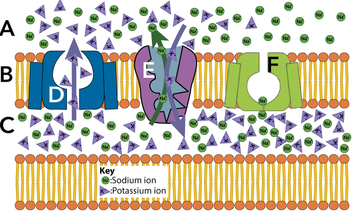
8. Nerve Impulse Conduction
The movement of a nerve impulse down the length of an axon is known as nerve impulse conduction or propagation. Let’s use what we’ve learned about action potentials to explain how nerve impulses move.
Here’s the same diagram we examined at the start of this tutorial (before we knew how action potentials worked). The diagram is a time sequence, showing a stretch of axon over a few milliseconds. In diagram A, the green patch at number 1 shows an area where an action potential is occurring: try to imagine the sodium ions diffusing into the cytoplasm through voltage gated channels. This flips the polarity of that stretch of neuron, causing it to become more positive on the inside than on the outside.
As these sodium ions enter, they’re causing the membrane potential of the adjacent region (A-2) to become less negative. At a certain point, so much Na+ has diffused through the membrane that the potential in region 2 rises above the threshold. This causes the voltage-gated sodium ion channels in that area to open, moving the action potential over to the right. This is represented in diagram B, where you can see the new action potential (B-1). At the same time, area B-3 shows a patch of membrane in the refractory period/overshoot.
Diagram C shows the action potential moving further to the right (area C-1). Meanwhile, the sodium-potassium pump has restored the resting potential in area C-4, and area C-3 is in the refractory period. This process will continue until the impulse reaches the end of the axon.
[qwiz random = “true” qrecord_id=”sciencemusicvideosMeister1961-Nerve Impulse Conduction, M28″]
[h]Nerve Impulse Propagation: Interactive Diagram
[q labels = “right”]
[l]resting potential
[fx] No, that’s not correct. Please try again.
[f*] Great!
[l]repolarization
[fx] No, that’s not correct. Please try again.
[f*] Correct!
[l]action potential
[fx] No. Please try again.
[f*] Great!
[q labels = “right”]
[l]Action potential begins.
[fx] No, that’s not correct. Please try again.
[f*] Good!
[l]Adjacent membrane region depolarizes to threshold. Action potential spreads.
[fx] No. Please try again.
[f*] Great!
[l]Membrane is at resting potential.
[fx] No, that’s not correct. Please try again.
[f*] Correct!
[l]Potassium channels open leading to repolarization.
[fx] No, that’s not correct. Please try again.
[f*] Correct!
[/qwiz]
9. Saltatory Conduction
If you accidentally touch a hot frying pan, the quicker you move your finger away, the less injury you’ll experience. Speedy reflexes, in other words, are highly adaptive. Within the animal kingdom, there have been two strategies for increasing nerve impulse propagation.
- Invertebrates (mollusks, earthworms, arthropods, etc.) have wide diameter axons. Increased diameter decreases resistance to the flow of ions (in the same way as a thick wire provides less resistance to electrical current than a thin wire does).
- Vertebrates partially insulate their axons with a myelin sheath. This enables action potentials to jump from one exposed area of the axon to the next, a kind of impulse propagation that’s called saltatory conduction. If you speak Spanish, that’s easy to remember, because the verb “saltar” means “to jump.” With myelination, impulses can move at speeds of up to 150 meters/second, whereas unmyelinated axons conduct impulses at velocities ranging from 0.5 to 10 meters/second. (Neuroscience, 2nd edition)
The myelin sheath is created by accessory cells that wrap themselves around portions of the axon.
 Here’s what this looks like in the central nervous system. Cells called oligodendrocytes (E) wrap themselves around the axon (D). This creates a myelin sheath (G), which is broken by uninsulated areas (F) that are called Nodes of Ranvier (after their French discoverer, Louis-Antoine Ranvier).
 In the peripheral nervous system, the myelin sheath (5) is created by cells called Schwann cells (6), that wrap themselves around the axon. As with the central nervous system, the insulation created by Schwann cells is not continuous. The gaps (7) go by the same name: Nodes of Ranvier.
 Here’s another image of a peripheral nerve cell. With the axon (4) wrapped in Schwann cells (2), action potentials (3) jump from one node of Ranvier (5) to the next.
[qwiz random = “true” qrecord_id=”sciencemusicvideosMeister1961-Saltatory Conduction, M28″]
[h]Saltatory Conduction
[i]
[q]In the diagram below, an oligodendrocyte is at
[textentry single_char=”true”]
[c]RQ ==[Qq]
[f]V2F5IHRvIGdvISBUaGUgb2xpZ29kZW5kcm9jeXRlIGlzIGF0IEUu[Qq]
[c]ICo=[Qq]
[f]IE5vLiBJbiB0aGUgY2VudHJhbCBuZXJ2b3VzIHN5c3RlbSwgdGhlIG9saWdvZGVuZHJvY3l0ZSBpcyB0aGUgY2VsbCB0aGF0IGNyZWF0ZXMgdGhlIG15ZWxpbiBzaGVhdGggKHdoaWNoIGlzIHNob3duIGF0IEcpLiBJZiBHIGlzIHRoZSBzaGVhdGgsIHdoYXQmIzgyMTc7cyB0aGUgb25seSBjYW5kaWRhdGUgZm9yIGEgY2VsbCB0aGF0IG1pZ2h0IGNyZWF0ZSB0aGlzIHNoZWF0aD8=[Qq]
[q]In the diagram below, the myelin sheath is at
[textentry single_char=”true”]
[c]Rw ==[Qq]
[f]TmljZSBqb2IhIFRoZSBteWVsaW4gc2hlYXRoIGlzIGF0IEc=[Qq]
[c]ICo=[Qq]
[f]IE5vLiBUaGUgbXllbGluIHNoZWF0aCBpcyB0aGUgaW5zdWxhdGluZyBsYXllciBhcm91bmQgdGhlIGF4b24uIEhlcmUmIzgyMTc7cyBhIGhpbnQ6IGl0JiM4MjE3O3MgY3JlYXRlZCBieSB0aGUgb2xpZ29kZW5kcm9jeXRlIGF0IEUu[Qq]
[q]In the diagram below, a Node of Ranvier is at
[textentry single_char=”true”]
[c]Rg ==[Qq]
[f]RXhjZWxsZW50ISBBIG5vZGUgb2YgUmFudmllciBpcyBhdCBG[Qq]
[c]ICo=[Qq]
[f]IE5vLiBUaGUgbm9kZXMgYXJlIHVubXllbGluYXRlZCBhcmVhcyBvZiBheG9uLg==[Qq]
[q]In the diagram below, a Node of Ranvier is at
[textentry single_char=”true”]
[c]Nw ==[Qq]
[f]RXhjZWxsZW50ISBBIG5vZGUgb2YgUmFudmllciBpcyBhdCA3[Qq]
[c]ICo=[Qq]
[f]IE5vLiBUaGUgbm9kZXMgYXJlIHVubXllbGluYXRlZCBhcmVhcyBvZiBheG9uLg==[Qq]
[q]In the diagram below, a Schwann cell is at
[textentry single_char=”true”]
[c]Ng ==[Qq]
[f]V2F5IHRvIGdvISBUd28gU2Nod2FubiBjZWxscyBhcmUgc2hvd24gYXQgNi4=[Qq]
[c]ICo=[Qq]
[f]IE5vLiBUaGUgU2Nod2FubiBjZWxscyBhcmUgd2hhdCBtYWtlIHVwIHRoZSBteWVsaW4gc2hlYXRoLg==[Qq]
[q]In the diagram below, letter G is the ________ sheath
[hangman]
[c]bXllbGlu[Qq]
[q]In the diagram below, a number 6 is a ________ cell.
[hangman]
[c]U2Nod2Fubg==[Qq]
[q]In the diagram below, a Schwann cell is at
[textentry single_char=”true”]
[c]Mg ==[Qq]
[f]V2F5IHRvIGdvISBBIFNjaHdhbm4gY2VsbCBpcyBzaG93biBhdCAyLg==[Qq]
[c]ICo=[Qq]
[f]IE5vLiBUaGUgU2Nod2FubiBjZWxscyBhcmUgd2hhdCBtYWtlIHVwIHRoZSBteWVsaW4gc2hlYXRoLg==[Qq]
[q]In the diagram below, an action potential is shown at
[textentry single_char=”true”]
[c]Mw ==[Qq]
[f]V2F5IHRvIGdvISBBbiBhY3Rpb24gcG90ZW50aWFsIGlzIHNob3duIGF0IDMu[Qq]
[c]ICo=[Qq]
[f]IE5vLiBIZXJlJiM4MjE3O3MgYSBoaW50LiBEdXJpbmcgYSByZXN0aW5nIHBvdGVudGlhbCwgdGhlIGN5dG9wbGFzbSBpcyBtb3JlIG5lZ2F0aXZlIHRoYW4gdGhlIGV4dHJhY2VsbHVsYXIgZmx1aWQuIER1cmluZyBhbiBhY3Rpb24gcG90ZW50aWFsLCBhIHJlZ2lvbiBvZiB0aGUgbWVtYnJhbmUgdGVtcG9yYXJpbHkgcmV2ZXJzZXMgcG9sYXJpdHku[Qq]
[q]The diagram below is demonstrating __________ conduction.
[hangman]
[c]c2FsdGF0b3J5[Qq]
[q]The myelinated neuron shown below is only found in ___________ animals.
[hangman]
[c]dmVydGVicmF0ZQ==[Qq]
[q]Compared to unmyelinated neurons, myelinated neurons (like the one shown below) are able to conduct impulses much _________.
[hangman]
[c]ZmFzdGVy[Qq]
[q]In the axon of the neuron in the diagram below, an action potential would only be recorded at
[textentry single_char=”true”]
[c]Nw ==[Qq]
[f]TmljZSEgSW4gYSBteWVsaW5hdGVkIG5ldXJvbiwgYWN0aW9uIHBvdGVudGlhbHMgb25seSBvY2N1ciBhdCB0aGUgTm9kZXMgb2YgUmFudmllci4=[Qq]
[c]ICo=[Qq]
[f]IE5vLiBIZXJlJiM4MjE3O3MgYSBoaW50LiBUaGUga2V5IGlkZWEgYWJvdXQgbXllbGluYXRlZCBuZXVyb25zIGlzIHRoYXQgYWN0aW9uIHBvdGVudGlhbCBjYW4ganVtcCBmcm9tIG9uZSBhcmVhIG9mIHRoZSBheG9uIHRvIHRoZSBuZXh0LiBUaGV5IGp1bXAgZnJvbSBfX19fXyB0byBfX19fX18u[Qq]
[x][restart]
[/qwiz]
Links
- Synapses and Summation (the next tutorial in this series)
- Nervous system main menu