Looking for a student learning guide? It’s on the main menu for your course. Use the “Courses” menu above.
1. Introduction: Animal bodies need to provide their cells with an isotonic environment
We ended the last tutorial on blood glucose regulation by examining why one of the first diagnostic signs of diabetes is excessive urination and thirst. Let’s generalize this by looking at how the body regulates the osmotic composition of body fluids, particularly blood.
One way to describe the composition of body fluids is in terms of osmolarity, which is a measure of how concentrated the solutes are within a solution. Add more sugar and lemon juice to your lemonade, and you’ve increased its osmolarity. Add more water to your lemonade, and you’ve decreased its osmolarity.
There’s a lot of overlap between concepts related to osmolarity and the concepts related to osmosis that we learned about earlier in this course. If you’re comparing two solutions, then the hypertonic solution is also the one with higher osmolarity. Conversely, the hypotonic solution is the one with lower osmolarity. And along the same lines, you can use the terms hyperosmolar, iso-osmolar, and hypoosmolar pretty much interchangeably with hypertonic, isotonic, and hypotonic.
The osmolarity of body fluids is, especially for terrestrial animals, a homeostatic variable that’s carefully regulated. Just for review, label the images below, then read the text that follows.
[qwiz summary = “false” qrecord_id=”sciencemusicvideosMeister1961-M26, Osmolarity”]
[h]Cells in various osmotic environments
[i]
[q labels = “top”]The environment of these red blood cells must be _________ to the cells.
[l]isotonic
[fx] No, that’s not correct. Please try again.
[f*] Excellent!
[l]hypertonic
[fx] No, that’s not correct. Please try again.
[f*] Correct!
[l]hypotonic
[fx] No. Please try again.
[f*] Great!
[q labels = “top”]The cells below are ______________ to their environment
[l]iso-osmolar
[fx] No, that’s not correct. Please try again.
[f*] Correct!
[l]hyperosmolar
[fx] No, that’s not correct. Please try again.
[f*] Great!
[l]hypoosmolar
[fx] No. Please try again.
[f*] Correct!
[q] Note that for animal cells, being in the right osmotic environment is a life-or-death situation. That’s because animal cells in an [hangman] environment (as in diagram “1”) will lose water to that environment and shrivel, while animal cells in a(n) [hangman] environment (diagram “3”) will gain water, eventually bursting. In both cases, water will be moving into or out of cells by [hangman].
[c]aHlwZXJ0b25pYw==[Qq]
[c]aHlwb3Rvbmlj[Qq]
[c]b3Ntb3Npcw==[Qq]
[x] So, how does your body keep the blood, and thereby the fluids surrounding your cells, isotonic to your cells?
Please continue reading below.
[/qwiz]
2. Osmoregulation is controlled by the hypothalamus through the pituitary gland
Keeping your body fluids isotonic to body tissues is known as osmoregulation, and the key regulator of your blood’s homeostatic set point for osmoregulation is the hypothalamus, a part of the brain that we’ve already met in the context of thermoregulation.
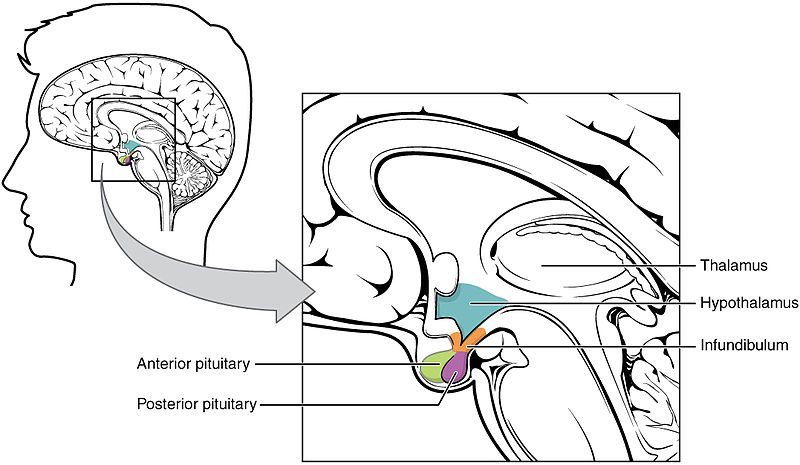
The hypothalamus is located just above the pituitary gland. When sensors in the hypothalamus detect that the blood has become too concentrated (hypertonic), they signal the pituitary to release the hormone ADH into the bloodstream.
3. ADH (Antidiuretic Hormone) Reduces Loss of Water in the Nephron’s Collecting Duct
ADH stands for antidiuretic hormone. A diuretic is a substance that increases the body’s excretion of urine. Diuretics are often used by doctors to treat high blood pressure (a condition that can lead to heart attacks, stroke, and organ damage). So let’s see how diuretics work, and then we can see how ADH works.
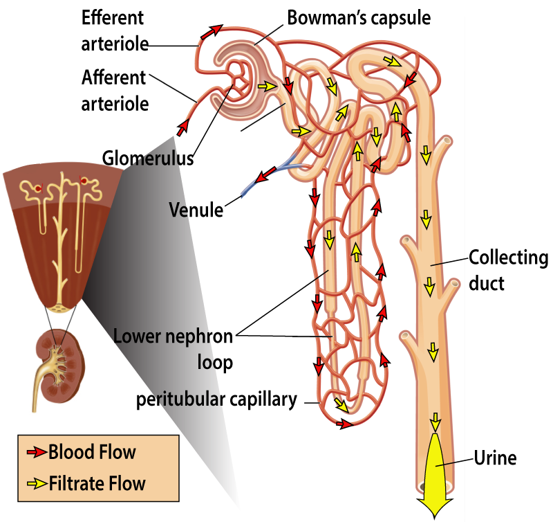
The diagram on the left shows the structure of the nephron tubule. Nephrons are the functional unit of the kidney, and there are about two million nephrons in each kidney. The afferent arteriole brings blood under pressure to a capillary bed called the glomerulus. The glomerulus acts as a filter, allowing a significant amount of the fluid in the blood to leak through and enter the nephron tubule as filtrate (which is the liquid that passes through a filter). You can follow the flow of this filtrate through the tubule by following the yellow arrows in the diagram).
The blood that doesn’t get filtered through the glomerulus enters the efferent arteriole, which subdivides into the peritubular capillaries. These capillaries, as their name indicates, surround the nephron tubule as it descends deep into the kidney, loops back up toward the kidney’s outer surface, and then descends again. You can follow the flow of the blood in the peritubular capillaries by looking at the red arrows.
The key point for understanding osmoregulation is that all along the length of the nephron tubule, fluid and solutes are exchanged between the filtrate in the tubule and the blood in the peritubular capillaries. This allows the kidneys to fine-tune the composition of the blood. The solutes and fluids that aren’t reabsorbed into the blood are excreted as urine.
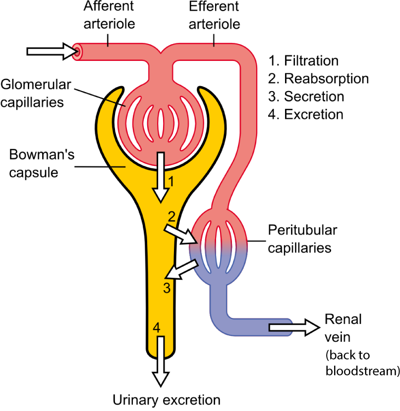
The diagram at right shows the same thing but in a much more schematic way. Using this diagram, let’s visualize how a diuretic works. Many diuretics increase the secretion of sodium ions from the peritubular capillaries into the nephron tubule (which in this diagram is everything below the capsule). The arrow at “3” would represent this flow of solute.
Now, adding solute makes the fluid in the tubule more hypertonic, causing water to flow from the peritubular capillaries into the tubule by osmosis. That increase in the amount of fluid in the tubule would, again, be represented by the arrow at “3.” With more fluid in the tubule, the amount of urine you excreted (at “4”) would increase. Because the kidney is excreting more water, it’s also decreasing the volume of water in the blood. That decreases blood pressure (because there’s less fluid in the blood vessels, which means less blood pushing out against the blood vessel walls). That, by the way, is why diuretics work as a blood pressure medication. Another effect will be to lower the solute concentration of the urine, which results in urine that’s lighter in color (because it’s more dilute, with relatively more water and less solute).
Now let’s look at ADH. Because it’s the antidiuretic hormone, ADH has the opposite effect that a diuretic has. ADH acts on the lower part of the tubule, a part of the nephron tubule called the collecting duct (see figure 3, above). ADH will bind to receptors on the cells that make up this part of the tubule and alter their membranes in a way that makes them more permeable to water. This causes water to leave the tubule, and to be reabsorbed back into the peritubular capillaries. Looking at Figure 4, imagine the arrow at “2” becoming larger ( indicating more reabsorption of fluid into the peritubular capillaries). As a result, there’s more solvent in the blood (where water is the solvent). The urine, by contrast, becomes more concentrated (darker), because it will have the same amount of solute, but less solvent.
Connecting osmoregulation with the color (and often the smell) of urine provides a very concrete way of understanding how osmoregulation works. If our body fluids are too hypotonic, our pituitary glands will release less ADH, and our kidneys will excrete urine that has more water and less solute. This is something that all of us have experienced every time we drink too much water. Our urine becomes copious (there’s a lot of it) and clear. Excretion of this watery urine reduces the amount of solvent in our body fluids, raising the osmolarity of our blood back to the set point. If our body fluids are too hypertonic (brought about by water loss caused by sweating a lot, or water deprivation), the hypothalamus will direct the pituitary glands to release ADH. As a result, our kidneys will try to maintain water, reabsorbing more water from the collecting duct and returning it to the peritubular capillaries. We’ll excrete as little water as possible, which means that our urine will be more concentrated: darker and probably smellier, too.
4. ADH and The Nephron Tubule: Checking Understanding Quiz 1
[qwiz qrecord_id=”sciencemusicvideosMeister1961-M26, ADH and the Nephron, 1″]
[h]ADH and the Nephron Tubule
[i]
[q] The regulatory process that works to keep body fluids isotonic to the cells is known as [hangman].
[c]IG9zbW9yZWd1bGF0aW9u[Qq]
[q] In the diagram below, the letter B represents the [hangman]. This part of the brain exerts regulatory control by influencing the secretions of the letter D, which represents the [hangman] gland.
[c]IGh5cG90aGFsYW11cw==[Qq]
[c]IHBpdHVpdGFyeQ==[Qq]
[q] One of the key hormones involved in regulating body fluid osmolarity is [hangman], which stands for anti[hangman] hormone
[c]IEFESA==[Qq]
[c]IGRpdXJldGlj[Qq]
[q] In the diagram below, the part that acts as a filter is shown at
[textentry single_char=”true”]
[c]ID I=[Qq]
[f]IEV4Y2VsbGVudC4gJiM4MjIwOzImIzgyMjE7IHNob3dzIHRoZSBnbG9tZXJ1bHVzLCB3aGljaCBhY3RzIGFzIGEgZmlsdGVyIGluIHRoZSBuZXBocm9uIHN5c3RlbQ==[Qq]
[c]IEVudGVyIHdvcmQ=[Qq]
[c]ICo=[Qq]
[f]IE5vLiBIZXJlJiM4MjE3O3MgYSBoaW50LiBXaGVuIHlvdSBtYWtlIGNvZmZlZSAoZnJvbSBncm91bmQgY29mZmVlKSwgdGhlIGZpcnN0IHRoaW5nIHlvdSBkbyBpcyBwb3VyIHdhdGVyIHRocm91Z2ggYSBmaWx0ZXIuIEluIHdoYXQmIzgyMTc7cyBhYm92ZSwgdGhlIGVxdWl2YWxlbnQgb2Ygd2F0ZXIgaXMgY29taW5nIGluIHRocm91Z2ggdGhlIA==YWZmZXJlbnQgYXJ0ZXJpb2xlLiBXaGVyZSBpcyB0aGUgYmxvb2QgZ29pbmcgYWZ0ZXIgdGhhdD8=[Qq]
[q] In the diagram below, the capsule that collects fluid so that it can enter the nephron tubule is shown at
[textentry single_char=”true”]
[c]ID E=[Qq]
[f]IE5pY2Ugam9iLiAmIzgyMjA7MSYjODIyMTsgc2hvd3MgdGhlIGNhcHN1bGUsIHdoaWNoIGNvbGxlY3RzIHRoZSBmbHVpZCB0aGF0IGZsb3dzIGludG8gdGhlIG5lcGhyb24gdHVidWxlLg==[Qq]
[c]IEVudGVyIHdvcmQ=[Qq]
[c]ICo=[Qq]
[f]IE5vLiBIZXJlJiM4MjE3O3MgYSBoaW50LiBUaGUgY2Fwc3VsZSBpcyByaWdodCBhZnRlciB0aGUgZ2xvbWVydWx1cywgd2hpY2ggaXMgYSBjYXBpbGxhcnkgYmVkIHRoYXQgeW91IGNhbiBmaW5kIHJpZ2h0IGFmdGVyIHRoZSBhZmZlcmVudCBhcnRlcmlvbGUu[Qq]
[q] In the diagram below, the nephron tubule is shown at
[textentry single_char=”true”]
[c]ID M=[Qq]
[f]IE5pY2Ugam9iLiAmIzgyMjA7MyYjODIyMTsgc2hvd3MgdGhlIG5lcGhyb24gdHVidWxlLg==[Qq]
[c]IEVudGVyIHdvcmQ=[Qq]
[c]ICo=[Qq]
[f]IE5vLiBIZXJlJiM4MjE3O3MgYSBoaW50LiBUaGUgeWVsbG93IGFycm93cyByZXByZXNlbnQgdGhlIGZsb3cgb2YgZmx1aWQgdGhyb3VnaCB0aGUgbmVwaHJvbiB0dWJ1bGUu[Qq]
[q] In the diagram below, the peritubular capillaries are shown at
[textentry single_char=”true”]
[c]ID Q=[Qq]
[f]IFRlcnJpZmljLiAmIzgyMjA7NCYjODIyMTsgc2hvd3MgdGhlIHBlcml0dWJ1bGFyIGNhcGlsbGFyaWVz[Qq]
[c]IEVudGVyIHdvcmQ=[Qq]
[c]ICo=[Qq]
[f]IE5vLiBIZXJlJiM4MjE3O3MgYSBoaW50LiBUaGUgcmVkIGFycm93cyByZXByZXNlbnQgdGhlIGZsb3cgb2YgYmxvb2QgdGhyb3VnaCB0aGUgcGVyaXR1YnVsYXIgY2FwaWxsYXJpZXMuIEFsc28sIHRoZSB3b3JkICYjODIyMDtwZXJpdHVidWxhciYjODIyMTsgbWVhbnMgJiM4MjIwO2Fyb3VuZCB0aGUgdHViZS4mIzgyMjE7[Qq]
[q] In the diagram below, the part of the nephron that ADH acts upon the most is
[textentry single_char=”true”]
[c]ID U=[Qq]
[f]IE5pY2Ugam9iLiAmIzgyMjA7NSYjODIyMTsgc2hvd3MgdGhlIGNvbGxlY3RpbmcgZHVjdCwgd2hpY2ggaXMgdGhlIG1haW4gdGFyZ2V0IG9mIEFESC4=[Qq]
[c]IEVudGVyIHdvcmQ=[Qq]
[c]ICo=[Qq]
[f]IE5vLiBIZXJlJiM4MjE3O3MgYSBoaW50LiBUaGUgbWFpbiB0YXJnZXQgb2YgQURIIGlzIHRoZSBwYXJ0IG9mIHRoZSB0dWJ1bGUgdGhhdCBkb2VzIHRoZSBmaW5hbCBwcm9jZXNzaW5nIG9mIHRoZSBmaWx0cmF0ZS4=[Qq]
[q] In the diagram below, filtration is represented by the number
[textentry single_char=”true”]
[c]ID E=[Qq]
[f]IE5pY2Ugam9iLiAmIzgyMjA7MSYjODIyMTsgcmVwcmVzZW50cyBmbHVpZCBiZWluZyBmaWx0ZXJlZCBvdXQgb2YgdGhlIGdsb21lcnVsdXMgYW5kIGVudGVyaW5nIHRoZSBjYXBzdWxlLg==[Qq]
[c]IEVudGVyIHdvcmQ=[Qq]
[c]ICo=[Qq]
[f]IE5vLiBIZXJlJiM4MjE3O3MgYSBoaW50LiAmIzgyMjA7QSYjODIyMTsgaXMgdGhlIGdsb21lcnVsdXMsIGEgbGVha3kgY2FwaWxsYXJ5IGJlZCB0aGF0IHNlbmRzIGZsdWlkIHRvIHBhcnQgJiM4MjIwO0IsJiM4MjIxOyB0aGUgY2Fwc3VsZS4gV2hhdCBudW1iZXIgaW5kaWNhdGVzIHRoZSBwcm9jZXNzIHRoYXQgaGFwcGVucyBiZXR3ZWVuICYjODIyMDtBJiM4MjIxOyBhbmQgJiM4MjIwO0I/JiM4MjIxOw==[Qq]
[q] In the diagram below, reabsorption is represented by number
[textentry single_char=”true”]
[c]ID I=[Qq]
[f]IEF3ZXNvbWUuICYjODIyMDsyJiM4MjIxOyBzaG93cyB0aGUgcmVhYnNvcnB0aW9uIG9mIGZsdWlkIGFzIGl0IG1vdmVzIGZyb20gdGhlIG5lcGhyb24gdHVidWxlICgmIzgyMjA7QyYjODIyMTspIGJhY2sgaW50byB0aGUgcGVyaXR1YnVsYXIgY2FwaWxsYXJpZXMgKCYjODIyMDtFJiM4MjIxOyku[Qq]
[q] In the diagram below, secretion is represented by number
[textentry single_char=”true”]
[c]ID M=[Qq]
[f]IEF3ZXNvbWUuICYjODIyMDszJiM4MjIxOyBzaG93cyBzZWNyZXRpb24gb2YgZmx1aWQgb3Igc29sdXRlIGFzIGl0IG1vdmVzIGZyb20gcGVyaXR1YnVsYXIgY2FwaWxsYXJpZXMgKCYjODIyMDtFJiM4MjIxOykgdG8gdGhlIG5lcGhyb24gdHVidWxlICgmIzgyMjA7QyYjODIyMTspLg==[Qq]
[c]IEVudGVyIHdvcmQ=[Qq]
[c]ICo=[Qq]
[f]IE5vLiBIZXJlJiM4MjE3O3MgYSBoaW50LiBTZWNyZXRpb24gaXMgd2hlbiBmbHVpZCBvciBzb2x1dGUgbW92ZXMgZnJvbSBwZXJpdHVidWxhciBjYXBpbGxhcmllcyAoJiM4MjIwO0UmIzgyMjE7KSB0byB0aGUgbmVwaHJvbiB0dWJ1bGUgKCYjODIyMDtDJiM4MjIxOyku[Qq]
[q] In the diagram below, excretion is represented by number
[textentry single_char=”true”]
[c]IDQ7 IEQ=[Qq]
[f]IE5pY2Ugam9iLiBQcm9jZXNzICYjODIyMDs0JiM4MjIxOyBzaG93cyB0aGUgZXhjcmV0aW9uIG9mIHdhc3RlcyBmcm9tIHRoZSB0dWJ1bGUgKHJlcHJlc2VudGVkIGJ5ICYjODIyMDtELCYjODIyMTsgd2hpY2ggaXMgYWxzbyBhbiBhY2NlcHRhYmxlIGFuc3dlciku[Qq]
[c]IEVudGVyIHdvcmQ=[Qq]
[c]ICo=[Qq]
[f]IE5vLiBIZXJlJiM4MjE3O3MgYSBoaW50LiBFeGNyZXRpb24gaXMgd2hlbiBzdWJzdGFuY2VzIGFyZSByZW1vdmVkIGZyb20gdGhlIGJvZHksIHNvIGp1c3QgbG9vayBhdCB0aGUgZW5kIG9mIHRoZSB0dWJ1bGUu[Qq]
[q] In the diagram below, the peritubular capillaries are represented by the letter
[textentry single_char=”true”]
[c]IE U=[Qq]
[f]IE5pY2Ugam9iLiBUaGUgcGVyaXR1YnVsYXIgY2FwaWxsYXJpZXMgYXJlIHJlcHJlc2VudGVkIGJ5ICYjODIyMDtFLiYjODIyMTs=[Qq]
[c]IEVudGVyIHdvcmQ=[Qq]
[c]ICo=[Qq]
[f]IE5vLiBIZXJlJiM4MjE3O3MgYSBoaW50LiBQZXJpdHVidWxhciBtZWFucyAmIzgyMjA7YXJvdW5kIHRoZSB0dWJlLCYjODIyMTsgYW5kIGNhcGlsbGFyaWVzIGFyZSBzbWFsbCBibG9vZCB2ZXNzZWxzLiBXaGVyZSBkbyB5b3Ugc2VlIHNtYWxsIGJsb29kIHZlc3NlbHMgYXJvdW5kIHRoZSB0dWJlPw==[Qq]
[q]If you’re dehydrated, your hypothalamus will get your [hangman] gland to secrete [hangman]. This hormone will increase the amount of [hangman] of fluid from the collecting duct. This will cause your urine to be [hangman] concentrated: that means it will be [hangman] and probably smellier.
[c]cGl0dWl0YXJ5[Qq]
[c]QURI[Qq]
[c]cmVhYnNvcnB0aW9u[Qq]
[c]bW9yZQ==[Qq]
[c]ZGFya2Vy[Qq]
[q]If your body fluids become too hypotonic, your hypothalamus will get your [hangman] gland to stop secreting [hangman]. As a result, you’ll reabsorb [hangman] fluid from the collecting duct in the [hangman]. This will cause your urine to be less [hangman].
[c]cGl0dWl0YXJ5[Qq]
[c]QURI[Qq]
[c]bGVzcw==[Qq]
[c]bmVwaHJvbg==[Qq]
[c]Y29uY2VudHJhdGVk[Qq]
[q] In the diagram below, ADH acts most upon which part?
[textentry single_char=”true”]
[c]IE M=[Qq]
[f]IEF3ZXNvbWUuICYjODIyMDtDJiM4MjIxOyBzaG93cyB0aGUgY29sbGVjdGluZyBkdWN0LCB3aGljaCBpcyB0aGUgcHJpbWFyeSB0YXJnZXQgb2YgQURILg==[Qq]
[c]IEVudGVyIHdvcmQ=[Qq]
[c]ICo=[Qq]
[f]IE5vLiBIZXJlJiM4MjE3O3MgYSBoaW50LiBBREggd2luZHMgdXAgZGVjcmVhc2luZyB0aGUgYW1vdW50IG9mIHVyaW5lIHByb2R1Y2VkLiBUbyByZWR1Y2UgdGhlIGFtb3VudCBvZiB1cmluZSwgd2hhdCBwYXJ0IG9mIHRoZSBzeXN0ZW0gYWJvdmUgd291bGQgaGF2ZSB0byBiZSB0YXJnZXRlZD8=[Qq]
[q] In the diagram below, numbers represent processes that occur along the nephron tubule. ADH would have its biggest effect on the number
[textentry single_char=”true”]
[c]ID I=[Qq]
[f]IEF3ZXNvbWUuICYjODIyMDsyJiM4MjIxOyBzaG93cyByZWFic29ycHRpb24uIEFESCBpbmNyZWFzZXMgdGhlIHJlYWJzb3JwdGlvbiBvZiBmbHVpZCBmcm9tIHRoZSBjb2xsZWN0aW5nIGR1Y3Qu[Qq]
[c]IEVudGVyIHdvcmQ=[Qq]
[c]ICo=[Qq]
[f]IE5vLiBIZXJlJiM4MjE3O3MgYSBoaW50LiBBREggd2luZHMgdXAgZGVjcmVhc2luZyB0aGUgYW1vdW50IG9mIHVyaW5lIHByb2R1Y2VkLiBXaGF0IG51bWJlcmVkIGFycm93IHdvdWxkIGhhdmUgdG8gYmUgYmlnZ2VyIHRvIG1ha2UgdGhlIGFtb3VudCBvZiB1cmluZSAoYXQgRCkgc21hbGxlcj8=[Qq]
[/qwiz]
5. Some Final Points about Osmoregulation: Aquaporins and Thirst
We saw above how ADH helps maintain the blood’s osmolarity by decreasing the amount of urine a water-stressed animal produces. To decrease urine flow, urine has to have increased osmolarity. Increased osmolarity means more solutes/unit volume. That will make the urine darker and smellier, something we’ve all experienced when we’re short of water.
How does ADH work on a cellular level? ADH increases the permeability of the collecting duct to water by inducing the cells lining the duct to insert protein channels called aquaporins into their membranes (figure 6 below, image A).
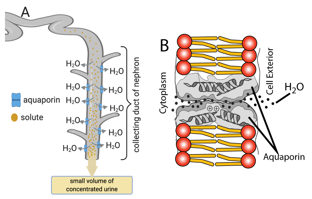
The aquaporins allow water to move by osmosis from inside the tubule to the outside of it while preventing the flow of solutes (see figure 6, image B). When ADH binds to receptors in the tubule, cellular changes cause vesicles with aquaporin subunits to move to the cells’ membranes. At the membrane, the subunits self-assemble into functional aquaporins, which allow water to leave the filtrate (from Biology: The Unity and Diversity of Life, Starr, Taggart,et. al, page 714).
Finally, let’s look at another effect of ADH that impacts the osmolarity of body fluids: it makes you thirsty. This makes sense: if your body fluids are becoming too hypertonic, one way to address that is to take in more solvent. The solvent in our bodies is water. Hence, when detectors in the hypothalamus detect low osmolarity, they directly send messages to other parts of the brain to generate feelings of thirst.
6. ADH and Osmoregulation: Cumulative Quiz
[qwiz qrecord_id=”sciencemusicvideosMeister1961-M26, ADH and Osmoregulation 2″]
[h]Osmoregulation: Cumulative Quiz
[i]
[q]In the diagram below, an increase in blood osmolarity would be detected by cells in the [hangman] (shown at “a”). This brain area would send messages that would generate a sensation of [hangman] (shown at “c”). Drinking water (at “i”), would [hangman] the osmolarity of the blood, bringing it back to the homeostatic set point.
[c]aHlwb3RoYWxhbXVz[Qq]
[c]dGhpcnN0[Qq]
[c]ZGVjcmVhc2U=[Qq]
[q]In the diagram below, the hormone [hangman] (at “d”), when it reaches the cells of the nephron’s [hangman] duct (at “e”), causes the cells in that area to allow [hangman] (at “f”) to diffuse into the [hangman] capillaries (at “g”). This keeps the [hangman] of the blood from increasing further.
[c]QURI[Qq]
[c]IGNvbGxlY3Rpbmc=[Qq]
[c]d2F0ZXI=[Qq]
[c]cGVyaXR1YnVsYXI=[Qq]
[c]b3Ntb2xhcml0eQ==[Qq]
[q] In the diagram below, ADH acts most upon which part?
[textentry single_char=”true”]
[c]IG U=[Qq]
[f]IEF3ZXNvbWUuICYjODIyMDtlJiM4MjIxOyBzaG93cyB0aGUgY29sbGVjdGluZyBkdWN0LCB3aGljaCBpcyB0aGUgcHJpbWFyeSB0YXJnZXQgb2YgQURILg==[Qq]
[c]IEVudGVyIHdvcmQ=[Qq]
[c]ICo=[Qq]
[f]IE5vLiBIZXJlJiM4MjE3O3MgYSBoaW50LiBBREggd2luZHMgdXAgZGVjcmVhc2luZyB0aGUgYW1vdW50IG9mIHVyaW5lIHByb2R1Y2VkLiBUbyByZWR1Y2UgdGhlIGFtb3VudCBvZiB1cmluZSwgd2hhdCBwYXJ0IG9mIHRoZSBzeXN0ZW0gYWJvdmUgd291bGQgaGF2ZSB0byBiZSB0YXJnZXRlZD8=[Qq]
[q] In the diagram below, fluid absorbed from the collecting duct reenters the blood at
[textentry single_char=”true”]
[c]IG c=[Qq]
[f]IE5pY2Ugam9iLiBMZXR0ZXIgJiM4MjIwO2cmIzgyMjE7IHNob3dzIHRoZSBwZXJpdHVidWxhciBjYXBpbGxhcmllcywgd2hpY2ggYWJzb3JiIHdhdGVyIGZyb20gdGhlIGNvbGxlY3RpbmcgZHVjdCwgcmV0dXJuaW5nIGl0IHRvIHRoZSBibG9vZHN0cmVhbS4=[Qq]
[c]IEVudGVyIHdvcmQ=[Qq]
[c]ICo=[Qq]
[f]IE5vLiBIZXJlJiM4MjE3O3MgYSBoaW50LiBBREggaW5kdWNlcyByZWFic29ycHRpb24gb2Ygd2F0ZXIgZnJvbSB0aGUgbmVwaHJvbiYjODIxNztzIGNvbGxlY3RpbmcgZHVjdCwgc2hvd24gYXQgJiM4MjIwO2YmIzgyMjE7LiBXaGF0IGRvZXMgdGhhdCB3YXRlciBnZXQgcmVhYnNvcmJlZCBpbnRvPw==[Qq]
[q] In the diagram below, what letter represents the pituitary gland?
[textentry single_char=”true”]
[c]IG I=[Qq]
[f]IEF3ZXNvbWUuIExldHRlciAmIzgyMjA7YiYjODIyMTsgc2hvd3MgdGhlIHBpdHVpdGFyeSBnbGFuZC4gVGhpcyBnbGFuZCByZWxlYXNlcyBBREggKGFzIHdlbGwgYXMgbWFueSBvdGhlciBob3Jtb25lcyku[Qq]
[c]IEVudGVyIHdvcmQ=[Qq]
[c]ICo=[Qq]
[f]IE5vLiBIZXJlJiM4MjE3O3MgYSBoaW50LiBUaGUgcGl0dWl0YXJ5IGdsYW5kIGlzIHRoZSBzb3VyY2Ugb2YgQURILCB3aGljaCBpcyBzaG93biBhdCAmIzgyMjA7ZC4mIzgyMjE7[Qq]
[q labels = “top”]
[l]darker
[fx] No, that’s not correct. Please try again.
[f*] Excellent!
[l]less
[fx] No. Please try again.
[f*] Good!
[l]lighter
[fx] No, that’s not correct. Please try again.
[f*] Correct!
[l]more
[fx] No, that’s not correct. Please try again.
[f*] Good!
[q]In the diagram below, the channels represented by “A” are [hangman].
[c]YXF1YXBvcmlucw==[Qq]
[q]A key idea represented by the diagram below is that [hangman] (represented by “A”) allow [hangman] to move out of the [hangman] duct while preventing the [hangman] of solutes (represented by “B”). The result is the excretion of highly concentrated [hangman], which reduces water loss.
[c]YXF1YXBvcmlucw==[Qq]
[c]d2F0ZXI=[Qq]
[c]Y29sbGVjdGluZw==[Qq]
[c]ZGlmZnVzaW9u[Qq]
[c]dXJpbmU=[Qq]
[q] In the diagram below, what letter represents increased secretion of ADH?
[textentry single_char=”true”]
[c]IE I=[Qq]
[f]IEF3ZXNvbWUuIExldHRlciAmIzgyMjA7QiYjODIyMTsgc2hvd3MgdGhlIHBpdHVpdGFyeSBnbGFuZCByZWxlYXNpbmcgQURILg==[Qq]
[c]IEVudGVyIHdvcmQ=[Qq]
[c]ICo=[Qq]
[f]IE5vLiBIZXJlJiM4MjE3O3MgYSBoaW50LiBUaGUgcGl0dWl0YXJ5IGdsYW5kIGlzIHRoZSBzb3VyY2Ugb2YgQURILg==[Qq]
[q] In the diagram below, what letter represents increased reabsorption of fluid from the collecting duct back into the bloodstream (note that the peritubular capillaries aren’t shown in this diagram: you have to imagine them).
[textentry single_char=”true”]
[c]IE M=[Qq]
[f]IE5pY2UuIExldHRlciAmIzgyMjA7QyYjODIyMTsgaXMgd2hlcmUgZmx1aWQgd291bGQgbGVhdmUgdGhlIGNvbGxlY3RpbmcgZHVjdCBhbmQgZ2V0IHJlYWJzb3JiZWQgYmFjayBpbnRvIHRoZSBibG9vZC4=[Qq]
[c]IEVudGVyIHdvcmQ=[Qq]
[c]ICo=[Qq]
[f]IE5vLiBIZXJlIGFyZSB0d28gaGludHMuMSkgVGhpcyB3b3VsZCBiZSBoYXBwZW5pbmcgYXQgdGhlIG5lcGhyb24mIzgyMTc7cyBjb2xsZWN0aW5nIGR1Y3QuIDIpIEl0JiM4MjE3O3MgYSByZXNwb25zZSB0byByaXNpbmcgb3Ntb2xhcml0eSwgYW5kIHRoZSBnb2FsIG9mIHRoZSByZXNwb25zZSBpcyB0byBsaW1pdCB0aGUgYW1vdW50IG9mIHdhdGVyIGxvc3QgZHVyaW5nIHRoZSBleGNyZXRpb24gb2YgdXJpbmUu[Qq]
[q] In the diagram below, what letter represents the excretion of hypertonic urine with relatively little water?
[textentry single_char=”true”]
[c]IE Q=[Qq]
[f]IFdheSB0byBnby4gTGV0dGVyICYjODIyMDtEJiM4MjIxOyByZXByZXNlbnRzIHRoZSBleGNyZXRpb24gb2YgaHlwZXJ0b25pYyB1cmluZS4=[Qq]
[c]IEVudGVyIHdvcmQ=[Qq]
[c]ICo=[Qq]
[f]IE5vLiBIZXJlJiM4MjE3O3MgYSBoaW50IChvciB0d28pLiAxKSBVcmluZSBpcyByZWxlYXNlZCBmcm9tIHRoZSBjb2xsZWN0aW5nIGR1Y3QuIDIpIElmIHlvdSYjODIxNztyZSBleGNyZXRpbmcgY29uY2VudHJhdGVkIChoeXBlcnRvbmljKSB1cmluZSwgaXQmIzgyMTc7cyBiZWNhdXNlIHlvdXIgYm9keSBpcyB0cnlpbmcgdG8gcHJlc2VydmUgd2F0ZXIsIHdoaWNoIGlzIHdoYXQmIzgyMTc7cyBkb25lIGluIHJlc3BvbnNlIHRvIHJpc2luZyBibG9vZCBvc21vbGFyaXR5Lg==[Qq]
[q] In the diagram below, what number represents the loop where “thirst” and “drinking” could be inserted as part of the response.
[textentry single_char=”true”]
[c]ID E=[Qq]
[f]IE5pY2Ugam9iLiBOdW1iZXIgMSByZXByZXNlbnRzIHRoZSBib2R5JiM4MjE3O3MgcmVzcG9uc2UgdG8gaGlnaCBvc21vbGFyaXR5LiBPbmUgd2F5IHRvIHJlZHVjZSBvc21vbGFyaXR5IHdvdWxkIGJlIHRvIHRha2UgaW4gbW9yZSB3YXRlci4=[Qq]
[c]IEVudGVyIHdvcmQ=[Qq]
[c]ICo=[Qq]
[f]IE5vLiBIZXJlJiM4MjE3O3MgYSBoaW50IChvciB0d28pLiBEcmlua2luZyB3YXRlciBhZGRzIHNvbHZlbnQgdG8geW91ciBib2R5IGZsdWlkcy4gSG93IHdvdWxkIHRoYXQgYWZmZWN0IG9zbW9sYXJpdHk/[Qq]
[q] In the diagram below, what letter would represent the release of a high volume of dilute urine.
[textentry single_char=”true”]
[c]IE g=[Qq]
[f]IE5pY2Ugam9iLiBMZXR0ZXIgSCByZXByZXNlbnRzIHdoYXQgd291bGQgZmxvdyBvdXQgb2YgdGhlIGNvbGxlY3RpbmcgZHVjdCBpbiByZXNwb25zZSB0byBmYWxsaW5nIGJsb29kIG9zbW9sYXJpdHkuIEZhbGxpbmcgb3Ntb2xhcml0eSBtZWFucyB0b28gbXVjaCB3YXRlciBpbiB0aGUgYmxvb2QsIGFuZCB0aGUgd2F5IHRoZSBib2R5IGNvdWxkIGdldCByaWQgb2YgdGhhdCB3b3VsZCBiZSB0byBleGNyZXRlIGEgbG90IG9mIGRpbHV0ZSB1cmluZS4=[Qq]
[c]IEVudGVyIHdvcmQ=[Qq]
[c]ICo=[Qq]
[f]IE5vLiBIZXJlJiM4MjE3O3MgYSBoaW50IChvciB0d28pLiAxLiBVcmluZSBpcyBnb2luZyB0byBjb21lIG91dCBvZiB0aGUgY29sbGVjdGluZyBkdWN0aW9uLiAyKSBFeGNyZXRpbmcgbG90cyBvZiBkaWx1dGUgdXJpbmUgbWVhbnMgdGhhdCB0aGUgYm9keSBpcyBnZXR0aW5nIHJpZCBvZiBzb2x2ZW50ICh3YXRlcikgdG8gaW5jcmVhc2UgYmxvb2Qgb3Ntb2xhcml0eS4gV2hlbiB3b3VsZCB0aGF0IGhhcHBlbj8=[Qq]
[x]
[restart]
[/qwiz]
Links
This tutorial ends (for now) with this module about animal homeostasis. At some point, I’d like to produce a tutorial on biological clocks. In the meanwhile:
- Click to return to the Feedback/ Homeostasis Main Menu
- Choose another module from the menu above