1. How do mRNA vaccines work?
Note: In the age of COVID, understanding immunity is a key part of scientific literacy. If you want to learn about the details of the immune system, you can do so through this Learn-Biology.com module. If you don’t have time for that, what’s below will give you enough background to understand mRNA vaccines: the most widely used type of vaccine against SARS-CoV-2.
When a person gets infected with a virus, their immune system launches a counterattack. That counterattack has many components, including
-
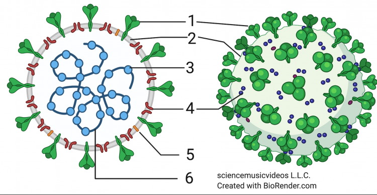
Antibodies (3) binding to and neutralizing a virus (1) Antibodies: Antibodies are proteins that bind with a virus. That neutralizes the virus and keeps it from entering and taking over cells.
- Killer T cells: These cells, also known as cytotoxic T cells, can find body cells that have been infected with the virus. The Killer T cells will kill these infected cells, keeping them from producing more viruses.
After the viral infection has been vanquished, you’ll develop immunity to that same virus. Immunity is based on
- Antibodies that continue to circulate in your bloodstream, which you’ll have for months following the initial infection.
- Memory cells. Memory cells are cells that your immune system creates following a successful counterattack against a virus (or any other pathogen). On a molecular level, memory cells “remember” the defeated virus, and wait in your lymph nodes, ready to counterattack if that same pathogen ever infects you again. The next time your body encounters that pathogen, your memory cells will quickly create antibodies and Killer T cells: so quickly that you’ll beat back the pathogen before you experience any symptoms.
A vaccine is a substance introduced into the body that trains your immune system to fight off a specific pathogen. Traditional vaccines (the ones we get as children against diseases like measles, mumps, polio, etc.) work by injecting either a weakened version of the virus, dead virus particles, or a small piece of that virus (usually a viral protein) into your body. What gets injected acts as an antigen, an “antibody generator.” In response to the antigen, your immune system creates antibodies and memory cells. This gives you immunity. If your body encounters that virus, you’ll quickly produce enough antibodies, killer T cells, and other immune system components to fight that virus off.
Vaccines have been in use for over 200 years. They prevent diseases that once caused millions of deaths (particularly among children) each year.
A variety of vaccines have been developed to provide us with immunity to SARS-CoV-2. You can learn more about these vaccines on this page on the NY Times website. For two reasons, I’m going to focus on one type of vaccine: mRNA vaccines. The first reason is that the way that mRNA vaccines work is particularly relevant to a college or AP-level Biology course. The second reason is that two leading vaccines used to protect against SARS-CoV-2 — the ones produced by the biotechnology companies Pfizer and Moderna — are mRNA vaccines. When you got vaccinated, these were most likely the vaccines that you received.
There are a few big ideas behind these mRNA vaccines:
- Unlike traditional vaccines, mRNA vaccines don’t use weakened or killed viruses (or even viral proteins) to elicit an immune response. Instead, they use a short segment of the virus’s mRNA. The mRNA used in the Pfizer and Moderna vaccines is a slightly modified version of the mRNA for the spike protein.
- Once injected, this mRNA goes into some of your cells, which start to produce the SARS-CoV-2 spike protein. The spike protein gets displayed on the membranes of these cells and is recognized by cells in the immune system.
- Recognition of the spike protein activates your immune system to learn how to recognize and fight off the SARS-CoV-2 virus.
Here’s some more detail about how mRNA vaccines work.
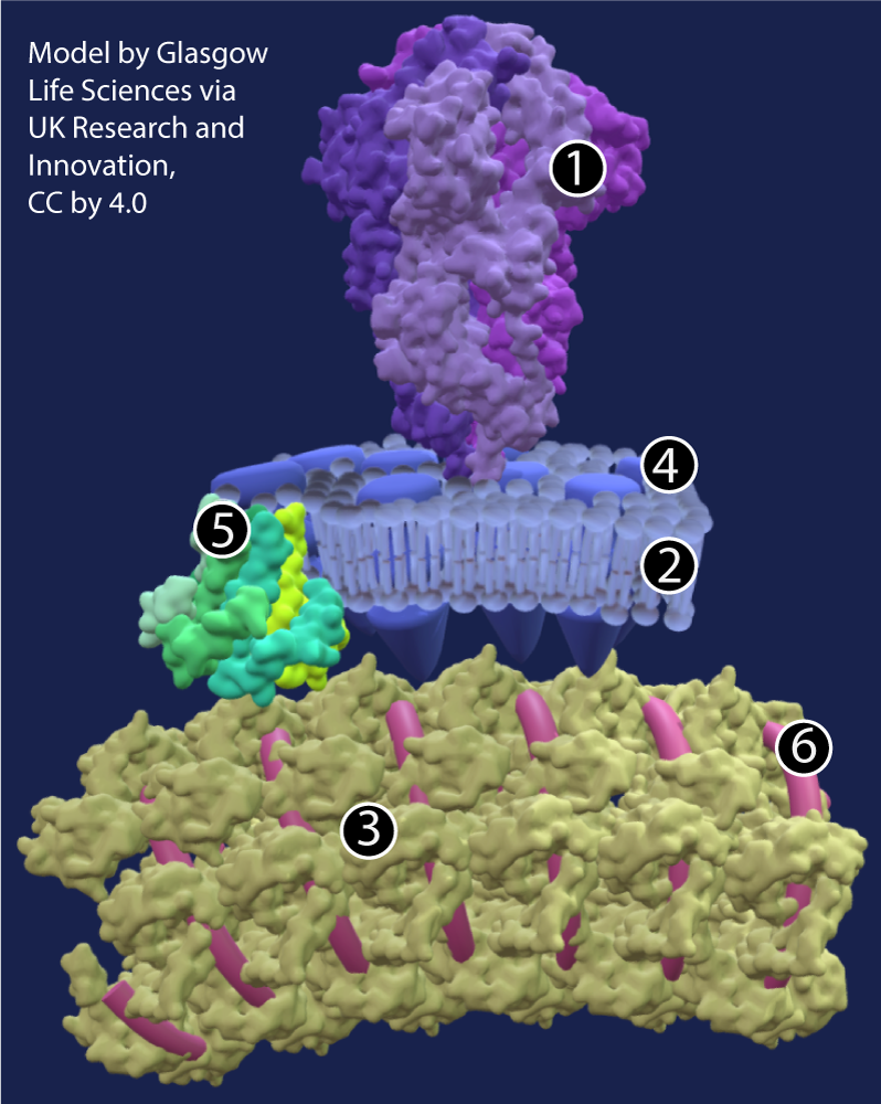
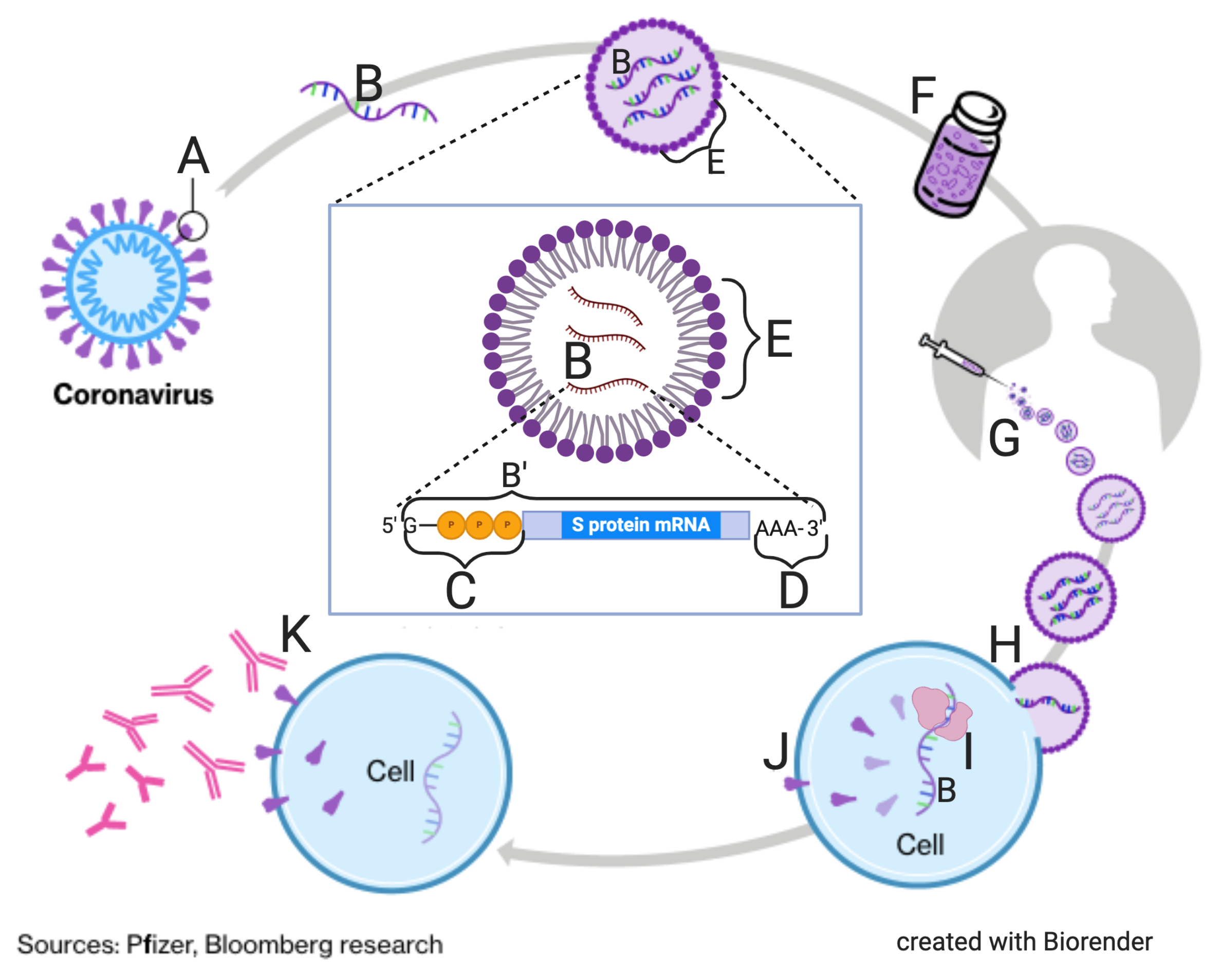 In the diagram to your left, “A” represents the spike protein of SARS-CoV-2. That protein was coded for by one of the protein-coding genes in the SARS-CoV-2 genome. “B” shows the RNA for that spike-protein-coding gene.
To make an mRNA vaccine, that mRNA is isolated, and modified in such a way that it’s ready for translation by a ribosome. This modification includes the addition of a 5′ GTP cap and a poly-A tail. Both of these modifications need to be engineered into the structure of an mRNA vaccine so that the mRNA can be translated by your ribosomes.
You can see this ready-to-translate mRNA shown at B’ in the large callout in the center of the image. At “C,” to the left of the S-protein mRNA sequence is a 5′ GTP cap. To the right, at the 3′ end of the mRNA is a poly-A tail (shown at “D”). The 5′ cap and the poly-A tail both serve to protect the mRNA from degradation by enzymes in the cytoplasm, giving the mRNA a long enough lifespan so that it can be translated by your cells.
Surrounding the mRNA is a lipid envelope, at E. This lipid envelope is what enables the viral mRNA to survive its trip through the bloodstream and enter a human cell.
Unlike DNA, which is a stable, tough molecule, mRNA is fragile. So is the lipid envelope. As a result, mRNA vaccines require ultra-cold storage. The Pfizer vaccine needs to be stored at -70° C. That requires a special freezer (much colder than the one you have in your kitchen). Moderna’s formulation is slightly less fragile, but still needs to be stored at -20° C (which is about the temperature of a kitchen freezer).
“F” shows a vaccine vial. Millions of these vials have been distributed around the U.S. (or any country where the public is getting vaccinated). “G” shows a hypodermic needle. A physician, nurse, or other trained technician injects you with the vaccine. The vaccine will float around in your bloodstream until it encounters one of your cells.
This encounter is shown at H. Your cell membranes are composed of lipids, and since lipids dissolve in lipids, the lipid envelope of the vaccine will fuse with your cells, dumping the viral mRNA inside.
Once inside your cells, the viral mRNA will be treated like any other mRNA. A ribosome (at “I”) will come along and read the viral mRNA. The result will be the production of the SARS-CoV-2 spike protein (at “J”). These proteins will then make their way to the surface of your cells.
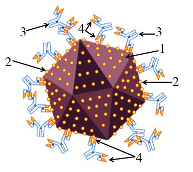
 At this point, I’m leaving out many steps. The key point, however, is that your immune system will identify this protein as an antigen. And once that happens, your immune system will be able to produce antibodies that will bind to the spike protein on the surface of SARS-CoV-2, as shown on the left. The antibodies are shown at 2. They’re binding to viral spike proteins, shown at 1. And once they do, the virus can no longer infect human cells. It’s been neutralized.
When your body knows how to do this, you have immunity. And thanks to mRNA vaccines you can get immunity without having to experience the symptoms associated with COVID-19.
2. mRNA vaccines: Checking Understanding
[qwiz random=”true” qrecord_id=”sciencemusicvideosMeister1961-mRNA vaccines (v2.0)”]
[h]Quiz: mRNA vaccines
[i]
[q] One goal of a vaccine is to get your [hangman] system to produce [hangman] cells. These cells enable your body to quickly respond to and fight off an infection.
[c]IGltbXVuZQ==[Qq]
[f]IEdyZWF0IQ==[Qq]
[c]IG1lbW9yeQ==[Qq]
[f]IEdyZWF0IQ==[Qq]
[q] Two vaccines that have been developed to fight COVID-19 both use the nucleic acid [hangman], enclosed within a( [hangman] envelope.
[c]IFJOQQ==[Qq]
[f]IEdvb2Qh[Qq]
[c]IGxpcGlk[Qq]
[f]IEdvb2Qh[Qq]
[q] The big idea behind mRNA vaccines is that they get your cells to produce SARS-CoV-2’s [hangman] protein (as shown in A and J). This enables your immune system to produce [hangman] to the virus (shown at K), enabling you to fight off subsequent infections.
[c]IHNwaWtl[Qq]
[f]IEV4Y2VsbGVudCE=[Qq]
[c]IGFudGlib2RpZXM=[Qq]
[f]IENvcnJlY3Qh[Qq]
[q] In the diagram below, mRNA coding for the spike protein is shown at
[textentry single_char=”true”]
[c]IE I=[Qq]
[f]IEV4Y2VsbGVudDogbVJOQSBmb3IgdGhlIHNwaWtlIHByb3RlaW4gaXMgc2hvd24gYXQgJiM4MjIwO0IuJiM4MjIxOw==[Qq]
[c]IEVudGVyIHdvcmQ=[Qq]
[c]ICo=[Qq]
[f]IE5vLiBIZXJlJiM4MjE3O3MgYSBoaW50LiBtUk5BIGlzIGEgc2luZ2xlLXN0cmFuZGVkIG51Y2xlaWMgYWNpZC4=[Qq]
[q] The goal of an mRNA vaccine is to get human cells to produce a viral protein, leading to the development of an immune response. In the diagram below, that human-produced viral protein is indicated by which letter?
[textentry single_char=”true”]
[c]IE o=[Qq]
[f]IE5pY2Ugam9iLiBBIGh1bWFuLXByb2R1Y2VkIHZpcmFsIHByb3RlaW4gaXMgc2hvd24gYXQgJiM4MjIwO0ouJiM4MjIxOw==[Qq]
[c]IEVudGVyIHdvcmQ=[Qq]
[c]ICo=[Qq]
[f]IE5vLiBIZXJlJiM4MjE3O3MgYSBoaW50LiBUaGVyZSYjODIxNztzIG9ubHkgb25lIHBsYWNlIGluIHRoZSBkaWFncmFtIHdoZXJlIHlvdSBzZWUgcHJvdGVpbiBzeW50aGVzaXMuIEZpbmQgdGhhdCBwbGFjZSwgYW5kIGl0JiM4MjE3O3Mgb25lIG9mIHRoZSBsZXR0ZXJzIGluIHRoYXQgcGFydCBvZiB0aGUgZGlhZ3JhbS4=[Qq]
[q] In the diagram below, the vaccine is shown fusing with a human cell at
[textentry single_char=”true”]
[c]IE g=[Qq]
[f]IEF3ZXNvbWUuIEggc2hvd3MgdGhlIHZhY2NpbmUgZnVzaW5nIHdpdGggYSBodW1hbiBjZWxsLg==[Qq]
[c]IEVudGVyIHdvcmQ=[Qq]
[c]ICo=[Qq]
[f]IE5vLiBIZXJlJiM4MjE3O3MgYSBoaW50LiBUaGlzIGZ1c2lvbiBiZXR3ZWVuIHRoZSB2YWNjaW5lIGFuZCBhIGh1bWFuIGNlbGwgbG9va3MgYSBiaXQgbGlrZSB0d28gc29hcCBidWJibGVzIGpvaW5pbmcgdG9nZXRoZXIu[Qq]
[q] In the diagram below, the lipid envelope that protects the injected mRNA when it’s in the bloodstream and which lets it get into a human cell is indicated by which letter?
[textentry single_char=”true”]
[c]IE U=[Qq]
[f]IEdvb2Qgd29yay4gTGV0dGVyICYjODIyMDtFJiM4MjIxOyBpcyB0aGUgbGlwaWQgZW52ZWxvcGUgdGhhdCBwcm90ZWN0cyB0aGUgbVJOQSBhbmQgd2hpY2ggbGV0cyBpdCBnZXQgaW50byBhIGh1bWFuIGNlbGwu[Qq]
[c]IEVudGVyIHdvcmQ=[Qq]
[c]ICo=[Qq]
[f]IE5vLiBIZXJlJiM4MjE3O3MgYSBoaW50LiBUaGUgbGlwaWQgZW52ZWxvcGUgc3Vycm91bmRzIHRoZSBtUk5BLg==[Qq]
[q] The goal of an mRNA vaccine is to get the immune system to produce antibodies to the virus In the diagram below, these antibodies are shown at
[textentry single_char=”true”]
[c]IE s=[Qq]
[f]IEF3ZXNvbWUuIEFudGlib2RpZXMgdG8gdGhlIHZpcnVzIGFyZSBzaG93biBhdCBLLg==[Qq]
[c]IEVudGVyIHdvcmQ=[Qq]
[c]ICo=[Qq]
[f]IE5vLiBIZXJlJiM4MjE3O3MgYSBoaW50LiBMb29rIGZvciBzb21ldGhpbmcgdGhhdCBzZWVtcyB0byBiZSByZXNwb25kaW5nIG9yIHJlYWN0aW5nIHRvIHRoZSBodW1hbi1wcm9kdWNlZCBzcGlrZSBwcm90ZWluIGF0ICYjODIyMDtKLiYjODIyMTs=[Qq]
[q] In the diagram below, a human ribosome is reading viral mRNA, producing a viral protein. That ribosome is shown at
[textentry single_char=”true”]
[c]IE k=[Qq]
[f]IEdvb2Qgd29yay4gJiM4MjIwO0kmIzgyMjE7IHNob3dzIGEgaHVtYW4gcmlib3NvbWUgcmVhZGluZyB2aXJhbCBtUk5BLg==[Qq]
[c]IEVudGVyIHdvcmQ=[Qq]
[c]ICo=[Qq]
[f]IE5vLiBIZXJlJiM4MjE3O3MgYSBoaW50LiBSaWJvc29tZXMgJiM4MjIwO3JlYWQmIzgyMjE7IG1STkEsIHByb2R1Y2luZyBwcm90ZWlucy4gRmluZCB0aGUgdmlyYWwgbVJOQSBpbnNpZGUgYSBodW1hbiBjZWxsLCBhbmQgeW91JiM4MjE3O2xsIGZpbmQgdGhlIGFuc3dlci4=[Qq]
[q] In the diagram below, a long string of repeated adenine nucleotides that protects the mRNA from enzymatic breakdown is shown at
[textentry single_char=”true”]
[c]IE Q=[Qq]
[f]IE5pY2Ugd29yay4gTGV0dGVyICYjODIyMDtEJiM4MjIxOyBzaG93cyB0aGUgcG9seS1BIHRhaWwsIGEgc3RyaW5nIG9mIHJlcGVhdGVkIGFkZW5pbmUgbnVjbGVvdGlkZXMgdGhhdCBwcm90ZWN0cyB0aGUgbVJOQSBmcm9tIGVuenltYXRpYyBhdHRhY2su[Qq]
[c]IEVudGVyIHdvcmQ=[Qq]
[c]ICo=[Qq]
[f]IE5vLiBIZXJlJiM4MjE3O3MgYSBoaW50LiBUaGlzIHBhcnQgb2YgdGhlIG1STkEgaXMgY2FsbGVkIGEgcG9seS1BIHRhaWwuIFdoYXQgb24gdGhlIGRpYWdyYW0gY291bGQgYmUgY2FsbGVkICYjODIyMDtwb2x5LUE/JiM4MjIxOw==[Qq]
[q]The diagram below shows a SARS-CoV-2 particle that’s been [hangman]. That’s because the [hangman] covering the spike proteins will keep the virus from [hangman] any more cells.
[c]bmV1dHJhbGl6ZWQ=[Qq]
[c]YW50aWJvZGllcw==[Qq]
[c]aW5mZWN0aW5n[Qq]
[/qwiz]
3. Introducing Rapid Antigen Tests
A rapid antigen test for SARS-CoV-2 tells you if the cells in your nose are producing a viral protein. If your test returns a positive result, it means that SARS-CoV-2 — the virus that causes COVID-19 — has made these cells into virus factories. That means that you have been made into a virus factory. You’re infectious. You need to isolate yourself so that you can avoid infecting others.
Please note two things about the material in this tutorial:
- what’s below is not intended as a guide to how to use a rapid antigen test kit, or even how to interpret the results. For that, please consult your test kit instructions. My goal is to teach the science behind how these tests work.
- As I’ll mention below, the same technology (lateral flow assay) is used to test for a variety of proteins found in human secretions. That includes a home pregnancy test, as well as tests for a variety of infections.
4. How Antibodies are Used to Detect Antigens in Rapid Tests
Let’s start with the name of the test.
- Rapid: They’re rapid because they return results in about 15 minutes. They can also be done at home. By comparison, the widely used PCR test has to be done in a lab, and receiving results can take hours or days.
- Antigen: “Antigen” means antibody generator. An antigen is a piece of some infectious agent (usually a virus or a bacterium) that causes your body to generate antibodies. In a COVID-19 rapid antigen test, the antigen is a protein that’s a part of SARS-CoV-2. If the test detects that antigen, it means that you’re infected and infectious.
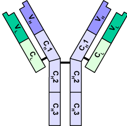
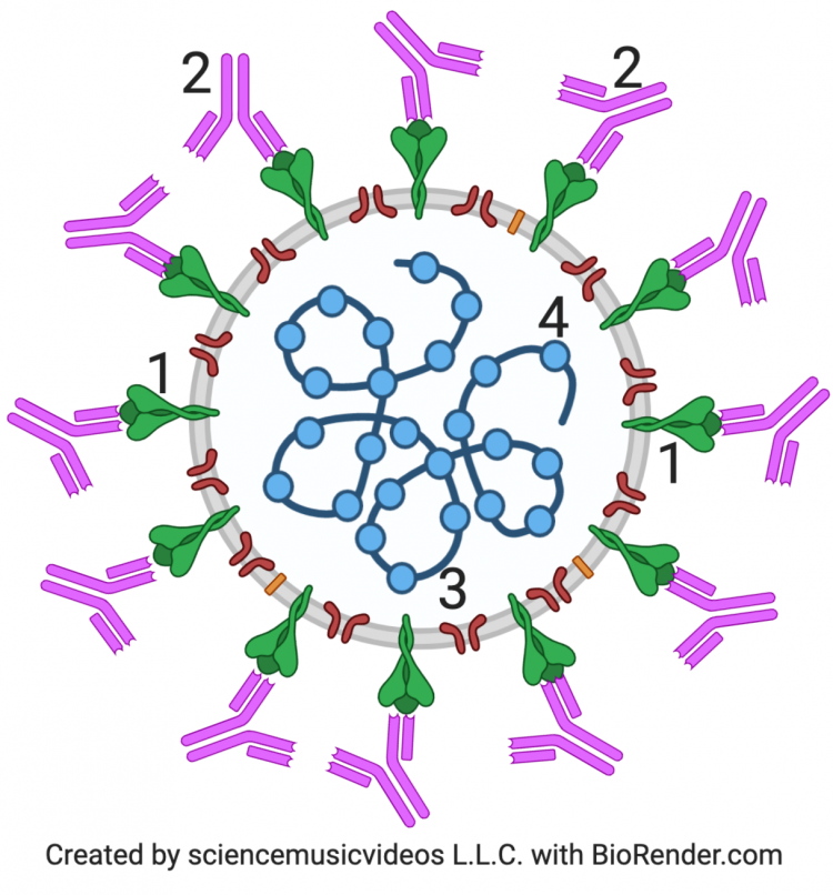
So what are antibodies? I introduced the term earlier in this module, and you can learn much more in this tutorial, but here’s a brief explanation. Antibodies are proteins that your immune system secretes to fight off pathogens. They have binding sites with very specific shapes, capable of binding to antigens, which either directly neutralize the infectious agent that brought the antigen into your body or act as a signal to your immune system to respond to the invader. In the diagram to the left, these binding sites are at the tips of the regions labeled with a “V.”

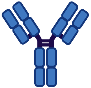
The image to the right is an illustration that gives an idea of what an antibody might look like if you shrunk yourself down to the size of a small molecule. Each bit of what looks like popcorn represents an atom. From this view, you can see the complexity of the shape at the tip of the “arms” of the antibody. That shape is an adaptation. It allows the antibody to bind with a specific antigen that has a complementary shape.
On the left, you see antibodies (in pink; two are labeled “2”) in action. The antibodies are binding to the spike protein of the coronavirus (which is shown in green). This is a key move in the body’s defense against the spike protein. That’s because once the antibody has bound itself to the spike protein antigen, SARS-CoV-2 is no longer able to infect you by entering cells.
In terms of testing, the important idea is that antibodies have a shape that enables them to bind with a specific antigen. In the rapid antigen tests that we’re learning about, antibodies that bind to a SARS-CoV-2 protein are used as the signal that SARS-CoV-2 is present (letting you know if you’re infectious).
The antigen that a COVID-19 rapid test detects is not the spike protein (“1” below in both diagrams). It’s the nucleocapsid protein (or N protein). The nucleocapsid protein is shown at “3” below. Its function is to surround and protect SARS-CoV-2’s RNA. If you’re infected with SARS-CoV-2, the virus will cause your cells to synthesize a lot of N protein. Therefore, detection of the N protein in your nasal secretions tells you that you’re infected and infectious.
 |
 |
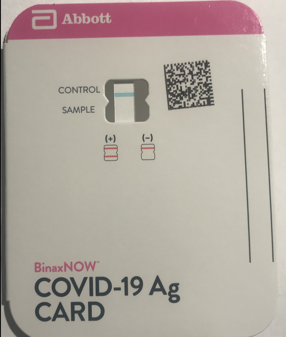
When you open your testing kit, what you’ll see will depend on the brand. Two widely available brands, Abbott and iHealth, are shown below. In the table below, you can see both what the kits look like, and what results they return.
| Abbott Laboratory’s BinaxNOW | iHealth COVID-19 Antigen Rapid Test |
Note the blue line that’s labeled “CONTROL.” |

“C” is for “control.” “T” is for “test.”Notice that the control line in the iHealth test only shows up after you carry out the test. |
| TEST RESULTS | |
 |
 |
The Abbott kit comes in a paper card, which you need to open to insert your sample. Inside the card is a test strip. All rapid-antigen kits have something like this inside (even though you might not be able to see it).
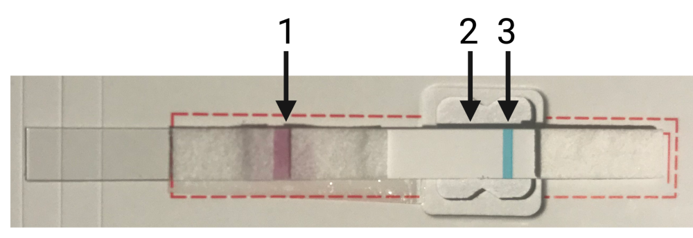
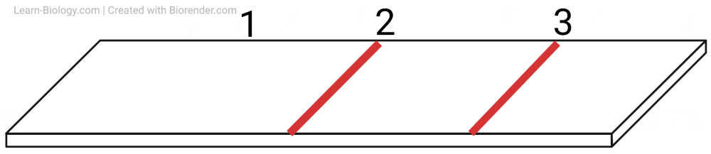
Number “3” is the control strip. If the test is working properly (whether or not you’re infected) the control strip will take on the purple color that you can see in line number 1. Arrow number 2 indicates where you’ll see a line for a positive test, or where you’ll see nothing if your test is negative. We’ll explain line number 1 below.
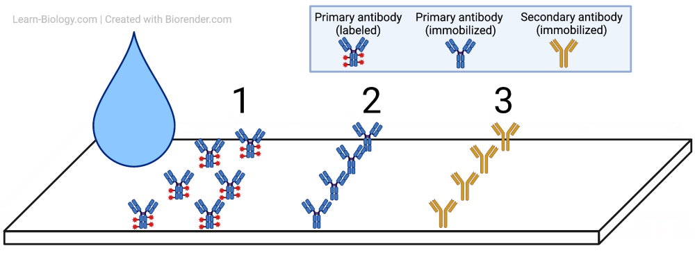
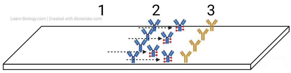
Let’s drop down to the molecular level. Note that the antibodies and viruses in the images below are not drawn to scale, but are drawn millions of times larger.
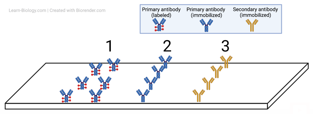

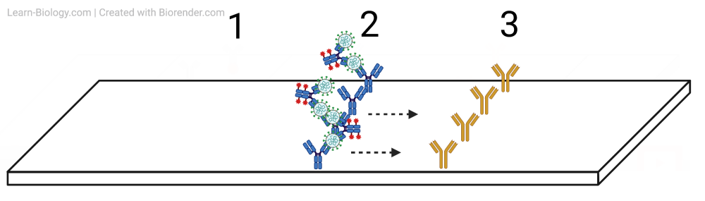
- Line 1 above corresponds to Line 1 on the test strip above. It contains labeled primary antibodies. What that means is that an antibody that binds with the target — the nucleocapsid protein — has been chemically engineered so that it’s now attached to another molecule. That other molecule (represented by the red star in the diagrams on the right, above, and below) is the label, and it lets us detect the presence of the labeled primary antibody. These antibody-label combinations are sitting on the (dry) test strip, but they’ll be able to move when the strip is wet, as we’ll see in a moment.
-
Unlabelled, immobilized primary antibody Line 2 consists of the same primary antibody, but with two differences. First, it hasn’t been labeled. Second, it’s been immobilized. It’s attached to that spot on the test strip.
-

Secondary antibody, immobilized Line 3 consists of a secondary antibody. It doesn’t bind with the N protein. Instead, it binds with the primary antibody. It’s also immobilized (attached to that spot on the test strip).
5. Positive results. You’re infectious (producing viral antigen)
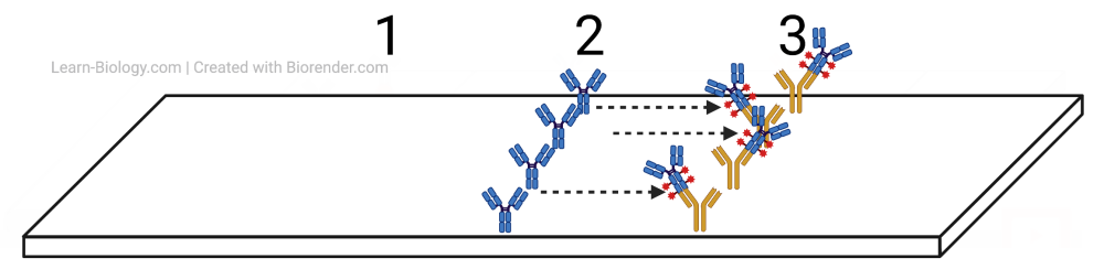
Now, imagine that you’ve swabbed your nose, collecting some of your nasal secretions. Imagine also that you’re infected with SARS-CoV-2. A droplet of your nasal secretions will contain millions of viruses, which you’re going to add to a designated spot on the left side of the test strip.

Once you add that droplet to the filter paper, the droplet (and everything in it) spreads across the paper through what’s called capillary action — just like a paper towel soaking up water. No pumping or gravity is required. The solution droplet, including your nasal secretions and any viruses that may be present, will spread across the length of the paper.
When that drop of virus-filled liquid reaches Line # 1, the N protein (the antigen) is going to bind with the labeled antibodies. Because of capillary action, those labeled antibodies, bound to viruses, are going to continue to move up the filter paper, towards lines 2 and 3.
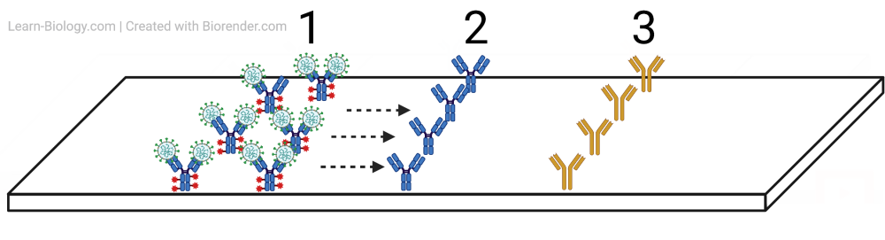
When those virus-bound labeled antibodies reach line # 2, they’ll bump into the immobilized primary antibodies. Because these immobilized antibodies at line # 2 also have binding sites for the N protein (the antigen), some of the labeled antibodies will get stuck at line # 2, as shown below.
On the test strip, those labeled antibodies will cause a signal (a red line) on the sample or test line that indicates the presence of the virus.
While millions of virus-bound labeled antibodies will get stuck at line 2, there will be some labeled antibodies that don’t bind with the virus. There will also be some virus-bound labeled antibodies that don’t bind to the antibodies at line 2. All of those antibodies, whether they have a virus attached or not, are going to continue to line 3, where they’ll bind with the immobilized secondary antibodies. Notice how the secondary antibodies aren’t binding with the virus; instead, they’re binding with the base of the primary antibodies.
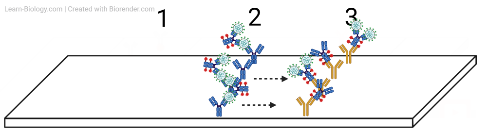
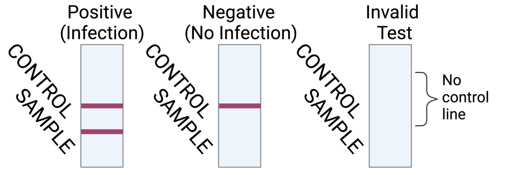
To shift away from the molecular view and to what you’ll observe when you take the test, the presence of viral antigen will lead to two lines:
The red line at “2” indicates the presence of the viral antigen (and therefore the virus). The line at 3 indicated that the test was working. That’s why a positive test has two lines.
6. Negative results. You’re not infectious (not producing viral antigen)
What if you’re not infectious? You’ve swabbed your nose, but there’s no virus in your nasal secretions.
In this case, the labeled primary antibodies will move up the filter paper, and pass right by the immobilized primary antibodies at line “2.” Remember, the primary antibodies at line “2” only bind to the virus’s protein, not to the other primary antibodies from line “1”. That’s why the line “1” antibodies keep on going.
When these labeled antibodies get to the control line, however, they’ll bind with the secondary antibodies. Again, the secondary antibodies at line “3” bind to the primary antibodies, whether or not they have viral protein.
This will cause the test to display a control signal, but not a sample signal.

7. An invalid result
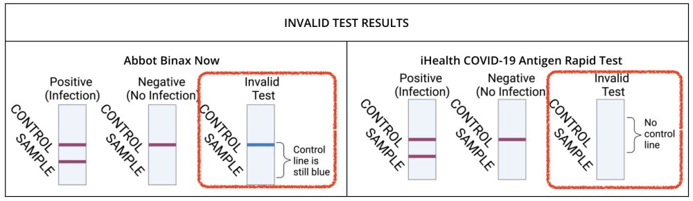
What if you don’t get the control line to change color (as with the Abbot Binax Now test) or to show up at all (as with the iHealth COVID-19 test)? In that case, the test is invalid. For some reason, you weren’t able to get the labeled primary antibodies to move across the test strip and bind with the immobilized secondary antibodies at the control line.
Re-read the instructions, and try again!
8. Rapid Antigen Tests: Checking Understanding
At this point, you should have a good understanding of how a rapid antigen test for COVID-19 works. While what’s above has focused on the detection of antigens from SARS-CoV-2, the same technology (called a lateral flow assay) is used in home pregnancy tests, as well as to test for strep throat infections, influenza, and malaria.
NOTE ABOUT ACCESSING THE QUIZ BELOW. As a public service, the quiz below is open to anyone. But if you’re in a course using Learn-Biology.com, you still need to sign in to get credit. Everyone else, just click “NO THANKS” when asked to log in or register.
[qwiz random=”true” qrecord_id=”sciencemusicvideosMeister1961-Rapid Antigen Tests (v.2.0)”]
[h]Rapid Antigen Tests: Checking Understanding
[i]
[q] A(n) [hangman] is a piece of a pathogen that causes your body’s immune system to launch its immune response.
[c]IGFudGlnZW4=[Qq]
[f]IEdvb2Qh[Qq]
[q] Antigen means “[hangman] generator.”
[c]IGFudGlib2R5[Qq]
[f]IEdyZWF0IQ==[Qq]
[q] In the diagram below, the antibody is at number
[textentry single_char=”true”]
[c]ID I=[Qq]
[f]IENvcnJlY3QhIE51bWJlciAyIHNob3dzIHRoZSBhbnRpYm9keS4=[Qq]
[c]IEVudGVyIHdvcmQ=[Qq]
[f]IFNvcnJ5LCB0aGF0JiM4MjE3O3Mgbm90IGNvcnJlY3Qu[Qq]
[c]ICo=[Qq]
[f]IEhlcmUmIzgyMTc7cyBhIGhpbnQuIEluIHRoaXMgZGlhZ3JhbSwgdGhlIGFudGlib2R5IGlzIGJpbmRpbmcgd2l0aCB0aGUgc3Bpa2UgcHJvdGVpbiBvZiB0aGUgY29yb25hdmlydXMu[Qq]
[q] In the diagram below, the nucleocapsid protein is shown at
[textentry single_char=”true”]
[c]ID M=[Qq]
[f]IE5pY2UhIFRoZSBudWNsZW9jYXBzaWQgcHJvdGVpbiAod2hpY2ggaXMgdGhlIGFudGlnZW4gaW4gdGhlIENPVklELTE5IHJhcGlkIHRlc3QpIGlzIHNob3duIGF0ICYjODIyMDszLiYjODIyMTs=[Qq]
[c]IEVudGVyIHdvcmQ=[Qq]
[f]IFNvcnJ5LCB0aGF0JiM4MjE3O3Mgbm90IGNvcnJlY3Qu[Qq]
[c]ICo=[Qq]
[f]IE5vLiBIZXJlJiM4MjE3O3MgYSBoaW50LiBUaGUgbnVjbGVvY2Fwc2lkIHByb3RlaW4gc3Vycm91bmRzIGFuZCBwcm90ZWN0cyB0aGUgdmlydXMmIzgyMTc7cyBSTkEuIFJOQSBpcyBhIGxvbmcgbW9sZWN1bGFyIHN0cmFuZC4gU28gZmluZCBzb21ldGhpbmcgdGhhdCYjODIxNztzIG9uIGEgbG9uZyBzdHJhbmQu[Qq]
[q] In the diagram below, the nucleocapsid protein is shown at
[textentry single_char=”true”]
[c]ID M=[Qq]
[f]IE5pY2UhIFRoZSBudWNsZW9jYXBzaWQgcHJvdGVpbiAod2hpY2ggaXMgdGhlIGFudGlnZW4gaW4gdGhlIENPVklELTE5IHJhcGlkIHRlc3QpIGlzIHNob3duIGF0ICYjODIyMDszLiYjODIyMTs=[Qq]
[c]IEVudGVyIHdvcmQ=[Qq]
[f]IFNvcnJ5LCB0aGF0JiM4MjE3O3Mgbm90IGNvcnJlY3Qu[Qq]
[c]ICo=[Qq]
[f]IE5vLiBIZXJlJiM4MjE3O3MgYSBoaW50LiBUaGUgbnVjbGVvY2Fwc2lkIHByb3RlaW4gc3Vycm91bmRzIGFuZCBwcm90ZWN0cyB0aGUgdmlydXMmIzgyMTc7cyBSTkEuIFJOQSBpcyByZXByZXNlbnRlZCBhcyBhIGxvbmcgY29pbC4=[Qq]
[q] On the test strip shown below, the primary labeled antibodies are found in which position?
[textentry single_char=”true”]
[c]ID E=[Qq]
[f]IFllcy4gUHJpbWFyeSBsYWJlbGVkIGFudGlib2RpZXMgYXJlIGZvdW5kIGF0IHBvc2l0aW9uICYjODIyMDsxLiYjODIyMTs=[Qq]
[c]IEVudGVyIHdvcmQ=[Qq]
[c]ICo=[Qq]
[f]IE5vLiBIZXJlJiM4MjE3O3MgYSBoaW50LiBJZiB0aGUgdGVzdCBpcyB2YWxpZCwgdGhlIHByaW1hcnkgbGFiZWxlZCBhbnRpYm9kaWVzIHdpbGwgY2F1c2UgdGhlIGNvbnRyb2wgbGluZSB0byBjaGFuZ2UgY29sb3IuIFdoYXQgY29sb3Igd2lsbCBpdCBjaGFuZ2UgdG8/IFdoZXJlIGRvIHlvdSBzZWUgdGhhdCBjb2xvcj8=[Qq]
[q] On the test strip shown below, immobilized primary unlabeled antibodies are found at which position?
[textentry single_char=”true”]
[c]ID I=[Qq]
[f]IFllcy4gSW1tb2JpbGl6ZWQgcHJpbWFyeSB1bmxhYmVsZWQgYW50aWJvZGllcyBhcmUgZm91bmQgYXQgcG9zaXRpb24gJiM4MjIwOzIuJiM4MjIxOw==[Qq]
[c]IEVudGVyIHdvcmQ=[Qq]
[c]ICo=[Qq]
[f]IE5vLiBIZXJlJiM4MjE3O3MgYSBoaW50LiBJZiB0aGUgdGVzdCBpcyBwb3NpdGl2ZSwgdGhlc2UgdW5sYWJlbGVkIGltbW9iaWxpemVkIHByaW1hcnkgYW50aWJvZGllcyB3aWxsIGJpbmQgd2l0aCB0aGUgdmlydXMgdGhhdCYjODIxNztzIGF0dGFjaGVkIHRvIHRoZSBwcmltYXJ5IGxhYmVsZWQgYW50aWJvZGllcy4gVGhhdCB3aWxsIHNob3cgdXAgYXMgYSBwaW5rL3JlZCBsaW5lIG9uIHRoZSB0ZXN0LiBXaGVyZSB3aWxsIHRoYXQgbGluZSBiZT8=[Qq]
[q] On the test strip shown below, immobilized secondary antibodies are found at line
[textentry single_char=”true”]
[c]ID M=[Qq]
[f]IFllcy4gSW1tb2JpbGl6ZWQgc2Vjb25kYXJ5IGFudGlib2RpZXMgYXJlIGZvdW5kIGF0IGxpbmUgJiM4MjIwOzMuJiM4MjIxOw==[Qq]
[c]IEVudGVyIHdvcmQ=[Qq]
[c]ICo=[Qq]
[f]IE5vLiBIZXJlJiM4MjE3O3MgYSBoaW50LiBUaGVzZSBpbW1vYmlsaXplZCBzZWNvbmRhcnkgYW50aWJvZGllcyBhcmUgZm91bmQgYXQgdGhlIGNvbnRyb2wgbGluZS4=[Qq]
[q labels = “top”]
[l]antigen
[fx] No, that’s not correct. Please try again.
[f*] Good!
[l]immobilized secondary antibody
[fx] No. Please try again.
[f*] Good!
[l]immobilized, unlabeled primary antibody
[fx] No. Please try again.
[f*] Great!
[l]primary labeled antibody
[fx] No, that’s not correct. Please try again.
[f*] Excellent!
[q multiple_choice=”true”]A color change (or the presence of a colored line, depending on your test) at position “3” in the diagram below indicates that
[c]IHRoZSBwcmVzZW5jZSBvZiBhbnRpZ2VuLg==[Qq]
[f]Tm8uIExpbmUgMyBpcyB0aGUgY29udHJvbCBsaW5lLiBTZWNvbmRhcnkgYW50aWJvZGllcyB3aWxsIGJpbmQgdG8gdGhlIHByaW1hcnksIGxhYmVsZWQgYW50aWJvZHkgYXQgdGhpcyBzcG90LiBUaGF0IG9ubHkgdGVsbHMgeW91IHRoYXQgdGhlIHRlc3QgaXMgdmFsaWQsIG5vdCB3aGV0aGVyIG9yIG5vdCB0aGVyZSYjODIxNztzIHZpcmFsIGFudGlnZW4u[Qq]
[c]IHRoZSBwcmltYXJ5IGxhYmVsZWQgYW50aWJvZHkgaGFzIGJvdW5k IHdpdGggaW1tb2JpbGl6ZWQgc2Vjb25kYXJ5IGFudGlib2RpZXM=[Qq]
[f]WWVzLiBXaGVuIHRoZSBwcmltYXJ5IGxhYmVsZWQgYW50aWJvZHkgKG9yaWdpbmFsbHkgYXQgcG9zaXRpb24gMSkgYmluZHMgd2l0aCB0aGUgaW1tb2JpbGl6ZWQgc2Vjb25kYXJ5IGFudGlib2RpZXMgYXQgbGluZSAzLCB0aGVyZSB3aWxsIGVpdGhlciBiZSBhIGNvbG9yIGNoYW5nZSAob3IgdGhlIGFwcGVhcmFuY2Ugb2YgYSBjb2xvcmVkIGxpbmUpLg==[Qq]
[c]IHRoZSBhYnNlbmNlIG9mIGFudGlnZW4=[Qq]
[f]Tm8uIExpbmUgMyBpcyB0aGUgY29udHJvbCBsaW5lLiBTZWNvbmRhcnkgYW50aWJvZGllcyB3aWxsIGJpbmQgdG8gdGhlIHByaW1hcnksIGxhYmVsZWQgYW50aWJvZHkgYXQgdGhpcyBzcG90LiBUaGF0IG9ubHkgdGVsbHMgeW91IHRoYXQgdGhlIHRlc3QgaXMgdmFsaWQsIG5vdCB3aGV0aGVyIG9yIG5vdCB0aGVyZSYjODIxNztzIGFudGlnZW4u[Qq]
[q labels=”top”]In a positive test, what happens in what order?
- _______________________________________________________________________________________________________________________________
- ______________________________________________________________________________________________________________________________
- ______________________________________________________________________________________________________________________________
[l]The primary labeled antibody binds with the secondary antibody.
[fx] No. Please try again.
[f*] Excellent!
[l]Antigen binds with the primary labeled antibody
[fx] No, that’s not correct. Please try again.
[f*] Correct!
[l]Antigen bound to the primary labeled antibody binds with the unlabeled, immobilized primary antibody.
[fx] No, that’s not correct. Please try again.
[f*] Excellent!
[q]The left side of the image below shows a rapid antigen test that’s returning a positive result. On a molecular level, what’s happening is that
[c]QW50aWdlbiBpcyBiaW5kaW5nIHdpdGggdGhlIGltbW9iaWxpemVkIHNlY29uZGFyeSBhbnRpYm9keQ==[Qq]
[f]Tm8uIFRoZSBpbW1vYmlsaXplZCBzZWNvbmRhcnkgYW50aWJvZHkgZG9lc24mIzgyMTc7dCBiaW5kIHdpdGggdGhlIGFudGlnZW4uIEl0IGJpbmRzIHdpdGggdGhlIHByaW1hcnkgYW50aWJvZHkuwqBIZXJlJiM4MjE3O3MgYSBoaW50OiB3aGF0JiM4MjE3O3MgaGFwcGVuaW5nIGJlbG93Pw==
Cg==[Qq]
[c]VGhlIHByaW1hcnkgbGFiZWxlZCBhbnRpYm9keSBpcyBiaW5kaW5nIHdpdGggdGhlIHNlY29uZGFyeSBhbnRpYm9keS4=[Qq]
[f]Tm8uIFRoYXQmIzgyMTc7cyB3aGF0IGhhcHBlbnMgYXQgdGhlIGNvbnRyb2wgbGluZS4gSGVyZSYjODIxNztzIGEgaGludDogd2hhdCYjODIxNztzIGhhcHBlbmluZyBiZWxvdz8=
Cg==[Qq]
[c]QW50aWdlbiBib3VuZCB0byB0aGUgcHJpbWFyeSBsYWJlbGVkIGFudGlib2R5IGJpbmRz IHdpdGggdGhlIHVubGFiZWxlZCwgaW1tb2JpbGl6ZWQgcHJpbWFyeSBhbnRpYm9keS4=[Qq]
[f]RXhjZWxsZW50LiBGb3IgdGhlcmUgdG8gYmUgYSBwb3NpdGl2ZSByZXN1bHQsIGFudGlnZW4gdGhhdCYjODIxNztzIGJvdW5kIHRvIHRoZSBwcmltYXJ5IGxhYmVsZWQgYW50aWJvZHkgaGFzIHRvIGJpbmQgd2l0aCB0aGUgdW5sYWJlbGVkLCBpbW1vYmlsaXplZCBwcmltYXJ5IGFudGlib2R5LCBhcyBzaG93biBiZWxvdzo=
Cg==[Qq]
[q] The control lines on the first and second images below indicate that the test is returning a valid result. On a molecular level, what’s happening is that
[c]QW50aWdlbiBpcyBiaW5kaW5nIHdpdGggdGhlIGltbW9iaWxpemVkIHNlY29uZGFyeSBhbnRpYm9keQ==[Qq]
[f]Tm8uIFRoZSBpbW1vYmlsaXplZCBzZWNvbmRhcnkgYW50aWJvZHkgZG9lc24mIzgyMTc7dCBiaW5kIHdpdGggdGhlIGFudGlnZW4uIEl0IGJpbmRzIHdpdGggdGhlIHByaW1hcnkgYW50aWJvZHkuwqBIZXJlJiM4MjE3O3MgYSBoaW50OiBMaW5lIDMgaXMgdGhlIGNvbnRyb2wgbGluZS4gV2hhdCYjODIxNztzIGhhcHBlbmluZz8=
Cg==[Qq]
[c]VGhlIHByaW1hcnkgbGFiZWxlZCBhbnRpYm9keSBiaW 5kcyB3aXRoIHRoZSBzZWNvbmRhcnkgYW50aWJvZHku[Qq]
[f]WWVzLiBJZiB0aGUgdGVzdCBpcyB2YWxpZCwgdGhhdCYjODIxNztzIHdoYXQgaGFwcGVucyBhdCB0aGUgY29udHJvbCBsaW5lLg==
Cg==[Qq]
[c]QW50aWdlbiBib3VuZCB0byB0aGUgcHJpbWFyeSBsYWJlbGVkIGFudGlib2R5IGJpbmRzIHdpdGggdGhlIHVubGFiZWxlZCwgaW1tb2JpbGl6ZWQgcHJpbWFyeSBhbnRpYm9keS4=[Qq]
[f]Tm8uIFRoYXQmIzgyMTc7cyB3aGF0IGhhcHBlbnMgaWYgdGhlcmUmIzgyMTc7cyBhIHBvc2l0aXZlIHJlc3VsdCAoaW5kaWNhdGVkIGJ5IHdoYXQmIzgyMTc7cyBoYXBwZW5pbmcgaW4gbGluZSAyLiBIZXJlJiM4MjE3O3MgYSBoaW50OiBMaW5lIDMgaXMgdGhlIGNvbnRyb2wgbGluZS4gV2hhdCYjODIxNztzIGhhcHBlbmluZz8=
Cg==[Qq]
[x]
[restart]
[/qwiz]
9. Exploring Further
There’s so much biology related to SARS-CoV-2— especially evolutionary biology — that I haven’t covered in this module. But that doesn’t mean that you can’t go on and learn it. I found these links to be particularly useful as I was researching this topic, and I encourage you to go ahead and explore.
- Coronavirus explained (links to multiple web pages, including “Getting to Know the New Coronavirus,” which has an excellent 3-D depiction of the virus)
- Sars-CoV-2 by the Numbers
- Wikipedia: Severe acute respiratory syndrome coronavirus 2
- HHMI Biointeractive, Biology of SARS-CoV-2
- Coronavirus Vaccine Tracker (NY times)
- How Pfizer’s [mRNA]Vaccine Works (NY Times)
- From Bats to Human Lungs, The Evolution of a Coronavirus (the New Yorker)
- A Primer on Coronavirus variants, mutation, and evolution (very good in the context of natural selection)
- A high school Q & A about Covid-19
- A Primer on and Conversation about the Biology and Evolution of SARS-CoV-2
- COVID-19 Variants
10. What’s next?
This tutorial ends this module about Viruses. The next tutorial in AP Bio Unit 6 is Topic 6.7, Part 4: Mutation.