1. Introducing cyclic AMP, the Second Messenger
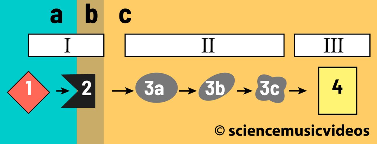
In the previous tutorials in this module, we learned that there are three phases involved in cell communication:
- I. Reception
- II. Signal transduction
- III. Cellular response.
We’ve also seen how a polar hormone such as epinephrine (represented by “1” in the diagram on your right) binds with a G-protein coupled receptor (“2”). Binding causes changes in the receptor, which in turn causes changes in a nearby G protein (“3”), allowing it to become activated by binding with GTP (“4”). This activated G-protein can now activate a nearby enzyme (“5”).
What follows (represented by “5” through “9”) is the focus of this tutorial. The movement of activity from the membrane to the cytoplasm involves transduction of the signal: changing the signal into a different form that induces cytoplasmic changes.
In our ongoing story of liver cells converting glycogen to glucose, the enzyme (represented by “5”) would be adenylyl cyclase. Adenylyl cyclase is widely shared among eukaryotes, and enzymes with similar functions are found among prokaryotes as well. The enzyme’s function is to convert ATP (represented by “6” above) into cyclic AMP (“7” above, and also on the right).

The “AMP” part of cyclic AMP stands for adenosine monophosphate. The single phosphate group in cyclic AMP is connected to the ribose sugar in the molecule with two covalent bonds). That’s why it’s called cyclic AMP, and it’s different in form and function from regular (non-cyclic) AMP. Cyclic AMP acts as a second messenger. Unlike the first messenger (epinephrine), cyclic AMP (or cAMP) goes into the cytoplasm, causing a series of changes that ultimately brings about a cellular response.
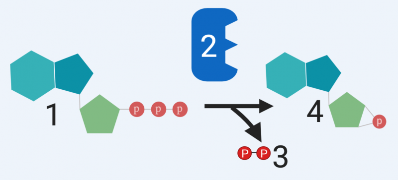 adenyl cyclase’s substrate, and two products result: two attached phosphate groups (at “3”) and cyclic AMP (at “4”)
After cAMP is created by adenylyl cyclase (“5” below), its role is to activate other proteins (represented by “8”), which then bring about the desired cellular response (“9”). What that means, in the context of our story in the liver, is activating “dormant” enzymes that break down the glycogen into glucose, enabling glucose to be released into the bloodstream to power the flight-or-flight response.
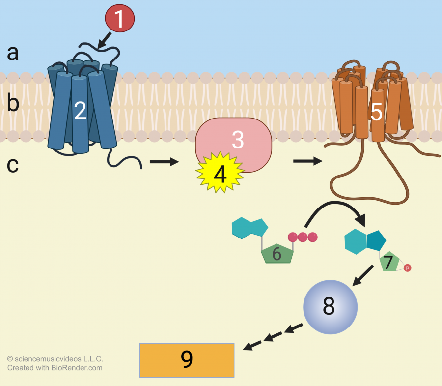 What this diagram doesn’t communicate, however, is that having second messengers like cAMP relay messages to other proteins allows for amplification of the original signal. Imagine a more complicated diagram in which adenylyl cyclase (“5”) is going to catalyze the production of many cAMPs (“7”), each of which will in turn activate many proteins (“8”) so that at the end of the process, millions of enzymes will have been activated.
In the context of epinephrine and the fight-or-flight response, each liver cell that receives the epinephrine signal will mobilize an enormous number of enzymes. As a result, within seconds glucose is coursing into the blood, on its way to your muscles.
I’ll explain the details of amplification in the next section. The last point, for now, is that as quickly as cAMP can be created from ATP, it can also be deactivated. This happens through an enzyme called phosphodiesterase (shown at “5” below). Phosphodiesterase converts cAMP (“4”) to plain old AMP (“6”). That deactivation of cAMP can shut down a fight-or-flight reaction as quickly as it got started. This is essential for survival: it would be maladaptive for you to continue to dump glucose into the blood when it’s not necessary. What makes evolutionary sense is to only break down glycogen when you need it.

2. Signal Transduction and cAMP
To consolidate what you’ve learned about signal transduction and cAMP, take the quiz below.
[qwiz random = “true” style=”width: 600px !important; min-height: 400px !important;” qrecord_id=”sciencemusicvideosMeister1961-signal transduction and cAMP (v2.0)”]
[h] Quiz: Signal Transduction and cAMP
[i]
[q json=”true” dataset_id=”SMV_Signal Transduction (Cell Communication)|6311ae923c7ff” question_number=”1″] In the diagram below, which number represents the ligand?
[textentry single_char=”true”]
[c]ID E=[Qq]
[f]IFllcy4g4oCcMeKAnSBpcyB0aGUgbGlnYW5kLCB0aGUgbW9sZWN1bGUgdGhhdCBiaW5kcyB3aXRoIGEgcmVjZXB0b3IgdG8gc2V0IG9mZiBhIGNlbGx1bGFyIHJlc3BvbnNlLg==[Qq]
[c]ICo=[Qq]
[f]IE5vLiBIZXJlJiM4MjE3O3MgYSBoaW50LiBBIGxpZ2FuZCBpcyBhIG1vbGVjdWxlIHRoYXQgYmluZHMgdG8gYW5vdGhlciAodXN1YWxseSBsYXJnZXIpIG1vbGVjdWxlLiBJbiB0aGlzIGRpYWdyYW0sIHdoYXQmIzgyMTc7cyBhYm91dCB0byBiaW5kIHdpdGggc29tZXRoaW5nIGxhcmdlcj8=
Cg==[Qq]
[q json=”true” dataset_id=”SMV_Signal Transduction (Cell Communication)|630c26f5fefff” question_number=”2″] In the diagram below, which number represents the receptor?
[textentry single_char=”true”]
[c]ID I=[Qq]
[f]IFllcy4g4oCcMuKAnSBpcyB0aGUgcmVjZXB0b3Iu[Qq]
[c]ICo=[Qq]
[f]IE5vLiBIZXJlJiM4MjE3O3MgYSBoaW50LiBUaGUgcmVjZXB0b3IgcmVjZWl2ZXMgYSBzaWduYWwgZnJvbSBvdXRzaWRlIHRoZSBjZWxsLiBJZiB0aGUgY29sb3IgYmx1ZSByZXByZXNlbnRzIHRoZSBjZWxsIGV4dGVyaW9yIGFuZCBiZWlnZSByZXByZXNlbnRzIHRoZSBjeXRvcGxhc20sIHRoZW4gd2hhdCYjODIxNztzIHRoZSBvbmx5IHRoaW5nIGluIHRoZSBtZW1icmFuZSB0aGF0IGlzIGFib3V0IHRvIGNvbm5lY3Qgd2l0aCBzb21ldGhpbmcgb3V0c2lkZSBvZiB0aGUgY2VsbD8=
Cg==[Qq]
[q json=”true” dataset_id=”SMV_Signal Transduction (Cell Communication)|63067a19033ff” question_number=”3″] In the diagram below, which number represents the G protein?
[textentry single_char=”true”]
[c]ID M=[Qq]
[f]IFllcy4g4oCcM+KAnSByZXByZXNlbnRzIHRoZSBHIHByb3RlaW4u[Qq]
[c]ICo=[Qq]
[f]IE5vLiBIZXJlJiM4MjE3O3MgYSBoaW50LiBUaGUgRyBwcm90ZWluIGdldHMgYWN0aXZhdGVkIGJ5IHRoZSByZWNlcHRvciAod2hpY2ggaXRzZWxmIGhhcyBiZWVuIGFjdGl2YXRlZCBieSB0aGUgbGlnYW5kKS4gVGhlbiB0aGUgRyBwcm90ZWluIGFjdGl2YXRlcyBhIG1lbWJyYW5lLWJvdW5kIGVuenltZSAoc2hvd24gYXQgJiM4MjIwOzUmIzgyMjE7KS4gV2hpY2ggaXMgdGhlIG9ubHkgcGFydCBvZiB0aGUgc3lzdGVtIGFib3ZlIHRoYXQgY291bGQgYmUgYWN0aXZhdGVkIGJ5IHRoZSByZWNlcHRvciwgYW5kIGluIHR1cm4sIGFjdGl2YXRlIHRoZSBtZW1icmFuZS1ib3VuZCBlbnp5bWUgYXQgJiM4MjIwOzU/JiM4MjIxOw==
Cg==[Qq]
[q json=”true” dataset_id=”SMV_Signal Transduction (Cell Communication)|6300f27cc5bff” question_number=”4″] In the diagram below, which number represents adenylyl cyclase (the enzyme that converts ATP to cyclic AMP)?
[textentry single_char=”true”]
[c]ID U=[Qq]
[f]IFllcy4g4oCcNeKAnSBpcyBhZGVueWx5bCBjeWNsYXNlLg==[Qq]
[c]ICo=[Qq]
[f]IE5vLiBIZXJlJiM4MjE3O3MgYSBoaW50LiBBZGVueWx5bCBjeWNsYXNlIGlzIGEgbWVtYnJhbmUtYm91bmQgZW56eW1lIHRoYXQgY29udmVydHMgQVRQIHRvIGN5Y2xpYyBBTVAuIFdoYXQmIzgyMTc7cyB0aGUgb25seSBwYXJ0IG9mIHRoZSBzeXN0ZW0gYWJvdmUgdGhhdCBsb29rcyBsaWtlIGEgbWVtYnJhbmUtYm91bmQgZW56eW1lIHRoYXQmIzgyMTc7cyBjb252ZXJ0aW5nIG9uZSB0aGluZyBpbnRvIGFub3RoZXIgdGhpbmc/
Cg==[Qq]
[q json=”true” dataset_id=”SMV_Signal Transduction (Cell Communication)|62f69d8801fff” question_number=”5″] In the diagram below, which number represents the second messenger cAMP?
[textentry single_char=”true”]
[c]ID c=[Qq]
[f]IFllcy4g4oCcN+KAnSByZXByZXNlbnRzIGNBTVAgKGN5Y2xpYyBBTVApIGEgc2Vjb25kIG1lc3Nlbmdlci4=[Qq]
[c]ICo=[Qq]
[f]IE5vLiBIZXJlJiM4MjE3O3MgYSBoaW50LiBDeWNsaWMgQU1QIGlzIHRoZSBzZWNvbmQgbWVzc2VuZ2VyLiBJdCB0YWtlcyB0aGUgaW5pdGlhbCBzaWduYWwgKGZyb20gdGhlIGxpZ2FuZCwgc2hvd24gYXQgJiM4MjIwOzEmIzgyMjE7KSBhbmQgdHJhbnNtaXRzIGl0IGludG8gdGhlIGN5dG9wbGFzbS4gV2hpY2ggcGFydCBvZiB0aGUgc3lzdGVtIGFib3ZlIGlzIHNlbmRpbmcgaW5mb3JtYXRpb24gZm9yd2FyZCBmcm9tIHRoZSBtZW1icmFuZSBpbnRvIHRoZSBjeXRvcGxhc20/
Cg==[Qq]
[q json=”true” dataset_id=”SMV_Signal Transduction (Cell Communication)|62f115ebc47ff” question_number=”6″] In the diagram below, which number represents the ATP that adenylyl cyclase converts into cAMP?
[textentry single_char=”true”]
[c]ID Y=[Qq]
[f]IFllcy4g4oCcNuKAnSBpcyB0aGUgQVRQIHRoYXQgdGhlIGFkZW55bHlsIGN5Y2xhc2UgZW56eW1lIChzaG93biBhdCAmIzgyMjA7NSYjODIyMTspIGNvbnZlcnRzIGludG8gY3ljbGljIEFNUC4=[Qq]
[c]ICo=[Qq]
[f]IE5vLiBIZXJlJiM4MjE3O3MgYSBoaW50LiBDeWNsaWMgQU1QIGlzIHRoZSBzZWNvbmQgbWVzc2VuZ2VyIHRoYXQgdHJhbnNtaXRzIGEgc2lnbmFsIHJlY2VpdmVkIGJ5IGEgcmVjZXB0b3IgaW4gdGhlIG1lbWJyYW5lIGludG8gdGhlIGN5dG9wbGFzbS4gQ3ljbGljIEFNUCBpcyBhIG1vZGlmaWVkIGZvcm0gb2YgQVRQLiBTZWUgaWYgeW91IGNhbiBpZGVudGlmeSB3aGljaCBtb2xlY3VsZSBpbiB0aGUgc3lzdGVtIGFib3ZlIGNvdWxkIHF1YWxpZnkgYXMgYSBzZWNvbmQgbWVzc2VuZ2VyLCBhbmQgdGhlbiBmaW5kIGl0cyBwcmVjdXJzb3IgZm9ybSAod2hpY2ggaXMgQVRQKS4=
Cg==[Qq]
[q json=”true” dataset_id=”SMV_Signal Transduction (Cell Communication)|62ebb390453ff” question_number=”7″] If the diagram below were about a liver cell being stimulated by the hormone epinephrine to produce glucose from glycogen, which number would represent epinephrine?
[textentry single_char=”true”]
[c]ID E=[Qq]
[f]IFllcy4g4oCcMeKAnSBpcyBlcGluZXBocmluZSwgdGhlIHNpZ25hbCB0aGF0IGJyaW5ncyBhYm91dCB0aGUgY2hhbmdlcyB0aGF0IGxlYWQgdGhpcyBsaXZlciBjZWxsIHRvIGNvbnZlcnQgaXRzIGdseWNvZ2VuIHN0b3JlcyBpbnRvIGdsdWNvc2Uu[Qq]
[c]ICo=[Qq]
[f]IE5vLiBIZXJlJiM4MjE3O3MgYSBoaW50LiBJZiBlcGluZXBocmluZSBpcyB0aGUgc2lnbmFsIHRoYXQgaXMgcmVjZWl2ZWQgYnkgdGhlIGNlbGwsIHdoYXQmIzgyMTc7cyB0aGUgb25seSBwYXJ0IG9mIHRoZSBzeXN0ZW0gYWJvdmUgdGhhdCBjb3VsZCByZXByZXNlbnQgdGhpcyBzaWduYWw/
Cg==[Qq]
[q json=”true” dataset_id=”SMV_Signal Transduction (Cell Communication)|62e67675843ff” question_number=”8″] If the diagram below were about a liver cell being stimulated by the hormone epinephrine to produce glucose from glycogen, which number would represent the conversion of glycogen into glucose?
[textentry single_char=”true”]
[c]ID k=[Qq]
[f]IFllcy4g4oCcOeKAnSBpcyB0aGUgY2VsbHVsYXIgcmVzcG9uc2UsIHdoaWNoIGluIHRoaXMgY2FzZSBpbnZvbHZlcyB0aGUgY2VsbCBjb252ZXJ0aW5nIGl0cyBnbHljb2dlbiBzdG9yZXMgaW50byBnbHVjb3NlLg==[Qq]
[c]ICo=[Qq]
[f]IE5vLiBIZXJlJiM4MjE3O3MgYSBoaW50LiBUaGUgcHJvZHVjdGlvbiBvZiBnbHVjb3NlIGZyb20gZ2x5Y29nZW4gaXMgdGhlIGNlbGx1bGFyIHJlc3BvbnNlLiBUaGUgcmVzcG9uc2UgaXMgdGhlIGxhc3QgdGhpbmcgdGhhdCBoYXBwZW5zIGluIHRoaXMgc3lzdGVtLg==
Cg==[Qq]
[q json=”true” dataset_id=”SMV_Signal Transduction (Cell Communication)|62e0c998887ff” question_number=”9″] In the diagram below, what number represents cyclic AMP (cAMP)?
[textentry single_char=”true”]
[c]ID Q=[Qq]
[f]IFllcy4g4oCcNOKAnSBpcyBjeWNsaWMgQU1QLCB0aGUgc2Vjb25kIG1lc3NlbmdlciBpbiBtYW55IHNpZ25hbCB0cmFuc2R1Y3Rpb24gc3lzdGVtcy4=[Qq]
[c]ICo=[Qq]
[f]IE5vLiBIZXJlJiM4MjE3O3MgYSBoaW50LiBDeWNsaWMgQU1QLCB0aGUgc2Vjb25kIG1lc3NlbmdlciBpbiBtYW55IGNlbGwgY29tbXVuaWNhdGlvbiBzeXN0ZW1zLCBpcyBtYWRlIGZyb20gQVRQIChhZGVub3NpbmUgdHJpcGhvc3BoYXRlKS4gU28gZmluZCBBVFAgKHdpdGggdGhyZWUgcGhvc3BoYXRlcykgYW5kIGZvbGxvdyB0aGUgZGlhZ3JhbSB0byBmaW5kIGNBTVAu
Cg==[Qq]
[q json=”true” dataset_id=”SMV_Signal Transduction (Cell Communication)|62db41fc4afff” question_number=”10″] In the diagram below, what number represents adenylyl cyclase?
[textentry single_char=”true”]
[c]ID I=[Qq]
[f]IFllcy4g4oCcMuKAnSBpcyBhZGVueWwgY3ljbGFzZSwgdGhlIGVuenltZSB0aGF0IGNyZWF0ZXMgdGhlIHNlY29uZCBtZXNzZW5nZXIgY3ljbGljIEFNUCBmcm9tIGl0cyBwcmVjdXJzb3IsIEFUUC4=[Qq]
[c]ICo=[Qq]
[f]IE5vLiBIZXJlJiM4MjE3O3MgYSBoaW50LiBGaW5kIHRoZSBlbnp5bWUgdGhhdCBjb252ZXJ0cyBBVFAgKHdpdGggdGhyZWUgcGhvc3BoYXRlcykgdG8gY3ljbGljIEFNUC4=
Cg==[Qq]
[q json=”true” dataset_id=”SMV_Signal Transduction (Cell Communication)|62d5dfa0cbbff” question_number=”11″] In the diagram below, what number represents phosphodiesterase?
[textentry single_char=”true”]
[c]ID U=[Qq]
[f]IFllcy4g4oCcNeKAnSBpcyBwaG9zcGhvZGllc3RlcmFzZSwgdGhlIGVuenltZSB0aGF0IGRlYWN0aXZhdGVzIGNBTVAgKCYjODIyMDs0JiM4MjIxOykgYnkgY29udmVydGluZyBpdCBpbnRvIEFNUCAoJiM4MjIwOzYmIzgyMjE7KQ==[Qq]
[c]ICo=[Qq]
[f]IE5vLiBIZXJlJiM4MjE3O3MgYSBoaW50LiBGaW5kIHRoZSBlbnp5bWUgdGhhdCBjb252ZXJ0cyBjeWNsaWMgQU1QIHRvIEFNUC4=[Qq]
[x][restart]
[/qwiz]
3. Signal Transduction through Phosphorylation Cascades Results in Signal Amplification
Now, let’s take a more detailed look at transduction, focusing on generating quick, short-term cytoplasmic responses. In the next tutorial, we’ll focus on longer-term changes that result from the activation of genes.
A key process that occurs as messages move from the membrane into the cytoplasm is phosphorylation, which is what happens when an enzyme adds a phosphate group to a molecule. You’ve seen phosphorylation before in the context of cellular respiration: it’s what happens when ADP receives a third phosphate group, creating ATP. It also happens (among other places) during the investment phase of glycolysis, when enzymes convert glucose into glucose-6-phosphate.
In the context of cell communication, enzymes called kinases phosphorylate proteins. In the same way that you can think of ATP as being more active than ADP, a phosphorylated protein can be thought of as the activated form of that protein.
Just to have a picture of what happens during phosphorylation, consider the image below:
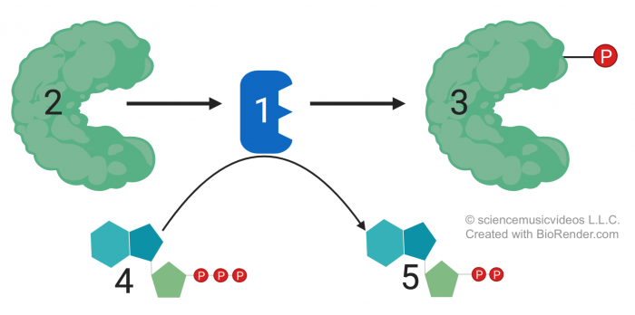 The enzyme at “1” is a kinase. It takes an unphosphorylated protein (at “2”) and adds a phosphate to it, converting it into a phosphorylated form (at “3”). The source of the phosphate is ATP (“4”), which becomes ADP (“5”) as a result of having lost a phosphate group.
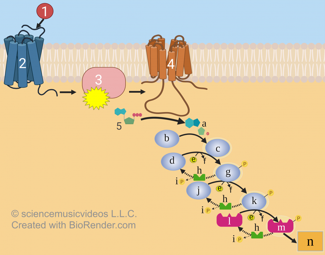
In cell communication, often the protein that gets phosphorylated by a kinase enzyme is itself a kinase. So what happens within a cell (like a liver cell) as it transduces a hormone signal into its cytoplasm is what’s called a phosphorylation cascade. cAMP gets the process started by activating the first kinase, and that one phosphorylates a second kinase, which activates a third, and so on. Here’s a diagram depicting the process.
You should already be familiar with 1 through 5 in the diagram, so we’ll start with “a,” which is cAMP. When cAMP contacts protein kinase “b,” that kinase is changed into its activated form, “c.” Because “c” is also a kinase, it will activate “d,” changing it into its active form, “g.” Note the little knob hanging off “g.” It’s a phosphate group. This phosphate group was donated by an ATP molecule, represented by “e.” The ADP that results is represented by “f.”
Because “g” is also a kinase, it will phosphorylate “j,” creating its activated form, “k.” Finally “k” will activate molecule “l,” which becomes “m.” In the case of epinephrine getting liver cells to break down glycogen and release glucose, you can imagine that “m” is the enzyme that does that. Letter “n” represents the cellular response, which is the production of glucose, which will diffuse out of these liver cells and into the bloodstream.
You might have noticed that there are still two parts of the diagram that we haven’t dealt with: the letters “h” and “i.” Letter “h” represents a type of enzyme called a protein phosphatase: these enzymes remove phosphates from kinases, deactivating them. As they do, they release a phosphate group into the cytoplasm, and that phosphate is represented by the letter “i.”
Now we can talk about signal amplification. Look again at the diagram, and imagine that cAMP isn’t just activating one molecule of the protein kinase represented by “b.” It’s converting many. Similarly, kinase “c” is converting many molecules of kinase “d” into “g.” The same is happening with “j” and “k” and with “l” and “m.”
So, when you create a mental image of a phosphorylation cascade, don’t imagine a single row of dominoes falling. Instead, think of an exponentially growing response. In the video below, Professor Chris Bishop has set up 100 ping pong balls on mouse traps. Each one of those traps is the equivalent of the dormant enzymes in a liver cell, waiting to be set off. Watch what happens in response to a small signal…a single ping pong ball.
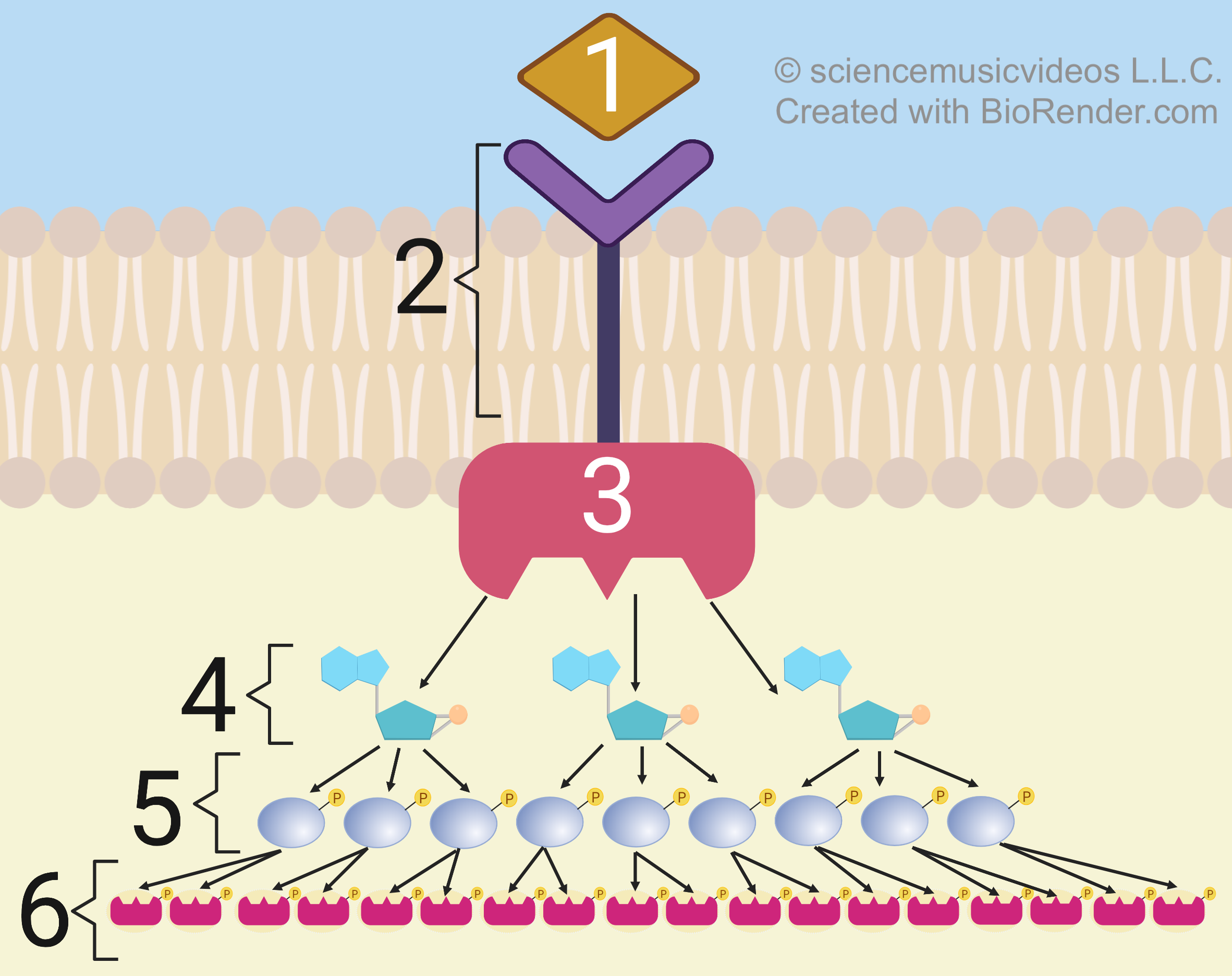
Here’s an image that’s trying to communicate the same idea:
The hormone (at “1)” binds with a membrane receptor (“2”), which activates a membrane-embedded enzyme (“3”) such as adenylyl cyclase. That enzyme activates a few second messengers (at “4”). Each second messenger will activate a few kinases (“5”), and each of these kinases will now activate multiple enzymes at the end of this chain (indicated by “6”).
One signal came in, and 18 enzymes have been activated as part of the cellular response.
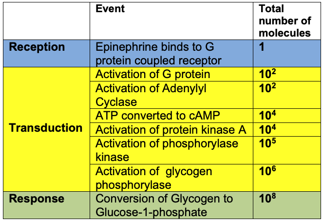
The graphic above, however, doesn’t nearly do justice to how amplified a cytoplasmic response can be. Study the diagram to your right. Notice that in each transition, the response might be amplified 10 or 100 times. A single epinephrine molecule might, in the end, be responsible for the release of 100 million glucose molecules…and that’s just one hormone binding to one receptor in one cell. Now think about the fact that there are hundreds of millions of liver cells, each of which has hundreds or thousands of receptors.
And that, of course, is also the reason why the cell needs to be able to turn these responses off as quickly as they were started. That’s why the cell has enzymes like phosphodiesterase to deactivate cAMP, and protein phosphatases to deactivate protein kinases.
While there are other systems to generate cytoplasmic responses, we’re going to limit this discussion to just G-protein coupled receptors, and the type of phosphorylation cascades we’ve just studied. In the next tutorial, we’ll address how other hormonal signals can generate longer-lasting, more permanent responses in cells by activating genes.
For now, let’s consolidate the learning we’ve done with a quiz.
4. Quiz: Kinases, Phosphorylation Cascades, and Signal Amplification
[qwiz random=”true” style=”width: 550px !important; min-height: 400px !important;” qrecord_id=”sciencemusicvideosMeister1961-Kinases, Phosphorylation Cascades, and Signal Amplification”]
[h] Kinases, Phosphorylation Cascades, and Signal Amplification
[i]
[q json=”true” dataset_id=”SMV_Kinases, Phosphorylation Cascades, Signal Amplification (cell communication)|62c15af8027ff” question_number=”1″] If molecule 2 is a protein, then which number represents a protein kinase?
[textentry single_char=”true”]
[c]ID E=[Qq]
[f]IFllcy4g4oCcMeKAnSBpcyBhIHByb3RlaW4ga2luYXNlLiBQcm90ZWluIGtpbmFzZXMgYXJlIGVuenltZXMgdGhhdCBwaG9zcGhvcnlsYXRlIHByb3RlaW5zLg==[Qq]
[c]ICo=[Qq]
[f]IE5vLiBIZXJlJiM4MjE3O3MgYSBoaW50LiBQcm90ZWluIGtpbmFzZXMgYXJlIGVuenltZXMgdGhhdCBwaG9zcGhvcnlsYXRlIHByb3RlaW5zIChhZGQgYSBwaG9zcGhhdGUgZ3JvdXAgdG8gdGhlbSwgd2hpY2ggYWx0ZXJzIHRoZWlyIGFjdGl2aXR5KS4gSW4gdGhlIGRpYWdyYW0gYWJvdmUsIHdoYXQgaXMgY29udmVydGluZyBhIHByb3RlaW4gaW50byBpdHMgcGhvc3Bob3J5bGF0ZWQgZm9ybT8=
Cg==[Qq]
[q json=”true” dataset_id=”SMV_Kinases, Phosphorylation Cascades, Signal Amplification (cell communication)|62ba5ed4567ff” question_number=”2″] In the diagram below, which number represents a phosphorylated protein?
[textentry single_char=”true”]
[c]ID M=[Qq]
[f]IFllcy4g4oCcM+KAnSBpcyBhIHByb3RlaW4gdGhhdCBoYXMgYmVlbiBwaG9zcGhvcnlsYXRlZCBieSBhIHByb3RlaW4ga2luYXNlIChzaG93biBhdCAmIzgyMjA7MSYjODIyMTspLiBZb3UgY2FuIHRlbGwgaXQmIzgyMTc7cyBiZWVuIHBob3NwaG9yeWxhdGVkIGJlY2F1c2Ugb2YgdGhlIGF0dGFjaGVkIHBob3NwaGF0ZSBncm91cCAodGhlICYjODIyMDtQJiM4MjIxOyBpbnNpZGUgYSB5ZWxsb3cgY2lyY2xlKS4=[Qq]
[c]ICo=[Qq]
[f]IE5vLiBIZXJlJiM4MjE3O3MgYSBoaW50LiBZb3UgY2FuIHRlbGwgaWYgYSBwcm90ZWluIGhhcyBiZWVuIHBob3NwaG9yeWxhdGVkIGlmIGl0IGhhcyBhIHBob3NwaGF0ZSBncm91cCBhdHRhY2hlZC4gVGFrZSBhIGdvb2QgbG9vayBhdCB0aGUgZGlhZ3JhbSBhYm92ZSwgYW5kIGZpbmQgdGhlIHBob3NwaG9yeWxhdGVkIHByb3RlaW4u
Cg==[Qq]
[q json=”true” dataset_id=”SMV_Kinases, Phosphorylation Cascades, Signal Amplification (cell communication)|62b25deb76bff” question_number=”3″] In the diagram below, which number represents the molecule that provided the phosphate that the protein kinase (1) used to phosphorylate molecule 2 into molecule 3?
[textentry single_char=”true”]
[c]ID Q=[Qq]
[f]IFllcy4g4oCcNOKAnSByZXByZXNlbnRzIEFUUC4gUHJvdGVpbiBraW5hc2VzIHRha2UgYSBwaG9zcGhhdGUgZnJvbSBBVFAgYW5kIHBsYW50IGl0IG9uIGFub3RoZXIgc3Vic3RyYXRlIG1vbGVjdWxlLCBwaG9zcGhvcnlsYXRpbmcgaXQu[Qq]
[c]ICo=[Qq]
[f]IE5vLiBIZXJlJiM4MjE3O3MgYSBoaW50LiBQcm90ZWluIGtpbmFzZXMgcGxhbnQgcGhvc3BoYXRlcyBvbnRvIGEgcHJvdGVpbi4gVG8gZG8gdGhhdCwgdGhleSBuZWVkIGEgcGhvc3BoYXRlICYjODIyMDtkb25vci4mIzgyMjE7IEZpbmQgYSBtb2xlY3VsZSB0aGF0IGxvc2VzIGEgcGhvc3BoYXRlIHNvIHRoYXQgYW5vdGhlciBtb2xlY3VsZSBjYW4gZ2FpbiBhIHBob3NwaGF0ZSwgYW5kIHlvdSYjODIxNztsbCBoYXZlIHRoZSBhbnN3ZXIu
Cg==[Qq]
[q json=”true” dataset_id=”SMV_Kinases, Phosphorylation Cascades, Signal Amplification (cell communication)|62ac668cfe7ff” question_number=”4″] In the diagram below, which letter represents cyclic AMP?
[textentry single_char=”true”]
[c]IG E=[Qq]
[f]IFllcy4gVGhlIGxldHRlciDigJxh4oCdIHJlcHJlc2VudHMgY3ljbGljIEFNUC4=[Qq]
[c]ICo=[Qq]
[f]IE5vLiBIZXJlJiM4MjE3O3MgYSBoaW50LiBDeWNsaWMgQU1QIGlzIHRoZSBzZWNvbmQgbWVzc2VuZ2VyIHRoYXQgbW92ZXMgdGhlIHNpZ25hbCBmcm9tIHRoZSBtZW1icmFuZSBpbnRvIHRoZSBjeXRvcGxhc20uIEluIHRoZSBkaWFncmFtIGFib3ZlLCB3aGljaCBpcyB0aGUgb25seSBtb2xlY3VsZSB0aGF0IGNvdWxkIGJlIHBsYXlpbmcgdGhhdCByb2xlPw==
Cg==[Qq]
[q json=”true” dataset_id=”SMV_Kinases, Phosphorylation Cascades, Signal Amplification (cell communication)|62a66f2e863ff” question_number=”5″] In the diagram below, which letter represents the inactive form of the first protein in this phosphorylation cascade?
[textentry single_char=”true”]
[c]IG I=[Qq]
[f]IFllcy4gTGV0dGVyIOKAnGLigJ0gcmVwcmVzZW50cyB0aGUgaW5hY3RpdmUgZm9ybSBvZiB0aGUgZmlyc3QgcHJvdGVpbiBpbiB0aGlzIHBob3NwaG9yeWxhdGlvbiBjYXNjYWRlLg==[Qq]
[c]ICo=[Qq]
[f]IE5vLiBIZXJlJiM4MjE3O3MgaG93IHRvIHRoaW5rIGFib3V0IHRoaXMuIExldHRlciAmIzgyMjA7YSYjODIyMTsgaXMgY0FNUCwgdGhlIHNlY29uZCBtZXNzZW5nZXIuIGNBTVAgYmluZHMgd2l0aCBhbiBpbmFjdGl2ZSBmb3JtIG9mIHRoZSBmaXJzdCBwcm90ZWluIGtpbmFzZSwgcmVzdWx0aW5nIGluIGEgcGhvc3Bob3J5bGF0ZWQgKHBob3NwaGF0ZS1jb250YWluaW5nKSB2ZXJzaW9uIG9mIHRoaXMgbW9sZWN1bGUgKHdoaWNoIHlvdSBjYW4gc2VlIGF0ICYjODIyMDtjJiM4MjIxOykuIExvb2sgYXQgdGhlIGRpYWdyYW0sIGFuZCB0cnkgdG8gZmlndXJlIG91dCB3aGF0IG1vbGVjdWxlIGNBTVAgKGF0ICYjODIyMDthJiM4MjIxOykgbXVzdCBiZSBpbnRlcmFjdGluZyB3aXRoIHRvIGJyaW5nIHRoaXMgYWJvdXQu
Cg==[Qq]
[q json=”true” dataset_id=”SMV_Kinases, Phosphorylation Cascades, Signal Amplification (cell communication)|62a02d4e917ff” question_number=”6″] In the diagram below, which letter represents the active form of protein kinase d?
[textentry single_char=”true”]
[c]IG c=[Qq]
[f]IFllcy4gTGV0dGVyIOKAnGfigJ0gcmVwcmVzZW50cyB0aGUgYWN0aXZlIGZvcm0gb2YgcHJvdGVpbiBraW5hc2UgZC4=[Qq]
[c]ICo=[Qq]
[f]IE5vLiBIZXJlJiM4MjE3O3MgaG93IHRvIHRoaW5rIGFib3V0IHRoaXMuIExldHRlciAmIzgyMjA7YSYjODIyMTsgaXMgY0FNUCwgdGhlIHNlY29uZCBtZXNzZW5nZXIuIGNBTVAgYmluZHMgd2l0aCBhbiBpbmFjdGl2ZSBmb3JtIG9mIHByb3RlaW4ga2luYXNlIGIsIHJlc3VsdGluZyBpbiBhIHBob3NwaG9yeWxhdGVkIChwaG9zcGhhdGUtY29udGFpbmluZykgdmVyc2lvbiBvZiB0aGlzIG1vbGVjdWxlOiB0aGUgYWN0aXZlIGZvcm0gd2hpY2ggeW91IGNhbiBzZWUgYXQgJiM4MjIwO2MmIzgyMjE7KS4gTG9vayBhdCB0aGUgZGlhZ3JhbSwgYW5kIHRyeSB0byBmaWd1cmUgb3V0IHdoYXQgbW9sZWN1bGUgbXVzdCBiZSB0aGUgYWN0aXZhdGVkIChwaG9zcGhvcnlsYXRlZCkgZm9ybSBvZiBwcm90ZWluIGtpbmFzZSBiLg==
Cg==[Qq]
[q json=”true” dataset_id=”SMV_Kinases, Phosphorylation Cascades, Signal Amplification (cell communication)|6299c62dde7ff” question_number=”7″] In the diagram below, which letter represents the enzyme that converts the phosphorylated kinases into their inactive forms?
[textentry single_char=”true”]
[c]IG g=[Qq]
[f]IFllcy4g4oCcaOKAnSByZXByZXNlbnRzIHRoZSBlbnp5bWUgKGEgcHJvdGVpbiBwaG9zcGhhdGFzZSkgdGhhdCB0cmFuc2Zvcm1zIHBob3NwaG9yeWxhdGVkIGtpbmFzZXMgKHN1Y2ggYXMgdGhvc2UgYXQgJiM4MjIwO2csJiM4MjIxOyBhbmQgJiM4MjIwO2ssJiM4MjIxOyB0byB0aGVpciB1bnBob3NwaG9yeWxhdGVkIGZvcm1zIChzdWNoIGFzIHRob3NlIGF0ICYjODIyMDtkJiM4MjIxOyBhbmQgJiM4MjIwO2ouJiM4MjIxOyk=[Qq]
[c]ICo=[Qq]
[f]IE5vLiBIZXJlJiM4MjE3O3MgaG93IHRvIHRoaW5rIGFib3V0IHRoaXMuIEZvY3VzIG9uIG1vbGVjdWxlICYjODIyMDtnLiYjODIyMTsgSXQmIzgyMTc7cyBhIHBob3NwaG9yeWxhdGVkIGtpbmFzZS4gTm90aWNlIHRoYXQgJiM4MjIwO2cmIzgyMjE7IHdhcyBtYWRlIGJ5IHBob3NwaG9yeWxhdGluZyBtb2xlY3VsZSAmIzgyMjA7ZCwmIzgyMjE7IGFuZCB0aGF0ICYjODIyMDtnJiM4MjIxOyBjYW4gYmUgY29udmVydGVkIGJhY2sgdG8gJiM4MjIwO2QuJiM4MjIxOyBCYXNlZCBvbiB3aGF0IHlvdSBzZWUgaW4gdGhlIGRpYWdyYW0sIHdoYXQmIzgyMTc7cyB0aGUgZW56eW1lIHRoYXQgY29udmVydHMgJiM4MjIwO2cmIzgyMjE7IGJhY2sgdG8gJiM4MjIwO2QmIzgyMjE7IChvciAmIzgyMjA7ayYjODIyMTsgYmFjayB0byAmIzgyMjA7aCYjODIyMTspPw==
Cg==[Qq]
[q json=”true” dataset_id=”SMV_Kinases, Phosphorylation Cascades, Signal Amplification (cell communication)|6293cecf663ff” question_number=”8″] In the diagram below, which letter represents the inactive form of the target protein that’s going to bring about the cellular response?
[textentry single_char=”true”]
[c]IG w=[Qq]
[f]IFllcy4gTGV0dGVyIOKAnGzigJ0gcmVwcmVzZW50cyB0aGUgaW5hY3RpdmUgKHVucGhvc3Bob3J5bGF0ZWQpIGZvcm0gb2YgdGhlIHRhcmdldCBwcm90ZWluLg==[Qq]
[c]ICo=[Qq]
[f]IE5vLiBIZXJlJiM4MjE3O3MgaG93IHRvIHRoaW5rIGFib3V0IHRoaXMuIFRoZSB1bHRpbWF0ZSBjZWxsdWxhciByZXNwb25zZSBpcyBpbmRpY2F0ZWQgYnkgdGhlIGxldHRlciAmIzgyMjA7bi4mIzgyMjE7IFRoaXMgY2VsbHVsYXIgcmVzcG9uc2UgaXMgYnJvdWdodCBhYm91dCBhdCB0aGUgZW5kIG9mIHRoaXMgcGhvc3Bob3J5bGF0aW9uIGNhc2NhZGUuIFdoYXQgbW9sZWN1bGUgbmVlZHMgdG8gYmUgcGhvc3Bob3J5bGF0ZWQgdG8gY3JlYXRlICYjODIyMDttPyYjODIyMTs=
Cg==[Qq]
[c]IEVudGVyIGxldHRlcg==[Qq]
[f]IFNvcnJ5LCBuby4=[Qq]
[q json=”true” dataset_id=”SMV_Kinases, Phosphorylation Cascades, Signal Amplification (cell communication)|628dd770edfff” question_number=”9″] In the diagram below, which letter represents the ATP that’s used during a phosphorylation cascade?
[textentry single_char=”true”]
[c]IG U=[Qq]
[f]IFllcy4gTGV0dGVyIOKAnGXigJ0gcmVwcmVzZW50cyBBVFAsIHRoZSBzb3VyY2Ugb2YgdGhlIHBob3NwaGF0ZSB1c2VkIHRvIHBob3NwaG9yeWxhdGUgdGhlIHByb3RlaW4ga2luYXNlcyBpbiB0aGlzIHBob3NwaG9yeWxhdGlvbiBjYXNjYWRlLg==[Qq]
[c]ICo=[Qq]
[f]IE5vLiBIZXJlJiM4MjE3O3MgaG93IHRvIHRoaW5rIGFib3V0IHRoaXMuIEluIHBob3NwaG9yeWxhdGlvbiBjYXNjYWRlcyBsaWtlIHRoZSBvbmUgcmVwcmVzZW50ZWQgYWJvdmUsIHByb3RlaW4ga2luYXNlcyBhY3RpdmF0ZSBvdGhlciBwcm90ZWlucyAod2hpY2ggY2FuIGFsc28gYmUga2luYXNlcykgYnkgYWRkaW5nIHBob3NwaGF0ZXMgdG8gdGhlbS4gVGhlIHBob3NwaGF0ZSBncm91cCBpcyB0YWtlbiBmcm9tIEFUUCwgd2hpY2ggYmVjb21lcyBBRFAgYXMgaXQgbG9zZXMgcGhvc3BoYXRlLiBTbyBsb29rIGF0IG1vbGVjdWxlcyAmIzgyMjA7ZCYjODIyMTsgYW5kICYjODIyMDtnLiYjODIyMTsgV2hhdCBzeW1ib2wgbGllcyBpbiBiZXR3ZWVuPyBZb3UmIzgyMTc7bGwgZmluZCB0aGUgc2FtZSBzeW1ib2wgYmV0d2VlbiAmIzgyMjA7aiYjODIyMTsgYW5kICYjODIyMDtrLCYjODIyMTsgYW5kIGV2ZW4gYmV0d2VlbiAmIzgyMjA7bCYjODIyMTsgYW5kICYjODIyMDttLiYjODIyMTs=
Cg==[Qq]
[q json=”true” dataset_id=”SMV_Kinases, Phosphorylation Cascades, Signal Amplification (cell communication)|6287e01275bff” question_number=”10″] Phosphorylation cascades like the one shown below need to be quickly activated, and also quickly deactivated. Which letter indicates a protein phosphatase, the enzyme that takes activated, phosphorylated molecules and deactivates them by removing a phosphate group?
[textentry single_char=”true”]
[c]IG g=[Qq]
[f]IFllcy4g4oCcaOKAnSByZXByZXNlbnRzIGEgcHJvdGVpbiBwaG9zcGhhdGFzZSwgdGhlIGRlYWN0aXZhdGluZyBlbnp5bWUgaW4gdGhpcyBwaG9zcGhvcnlsYXRpb24gY2FzY2FkZS4=[Qq]
[c]ICo=[Qq]
[f]IE5vLiBTZWUgaWYgeW91IGNhbiBmaW5kIGEgbGV0dGVyIHRoYXQgcmVwcmVzZW50cyBhbiBlbnp5bWUgdGhhdCB3b3VsZCBjb252ZXJ0IGEgcGhvc3Bob3J5bGF0ZWQga2luYXNlIHN1Y2ggYXMgJiM4MjIwO2cmIzgyMjE7IGJhY2sgaW50byBpdHMgdW5waG9zcGhvcnlsYXRlZCBmb3JtIGF0ICYjODIyMDtkLiYjODIyMTsgVGhlIHNhbWUgZW56eW1lIGlzIGF0IHdvcmsgYmV0d2VlbiAmIzgyMjA7ayYjODIyMTsgYW5kICYjODIyMDtqJiM4MjIxOyBvciAmIzgyMjA7bSYjODIyMTsgYW5kICYjODIyMDtsLiYjODIyMTs=
Cg==[Qq]
[q json=”true” dataset_id=”SMV_Kinases, Phosphorylation Cascades, Signal Amplification (cell communication)|6282333579fff” question_number=”11″] In the diagram below, the ligand is represented by which number?
[textentry single_char=”true”]
[c]ID E=[Qq]
[f]IFllcy4g4oCcMeKAnSB0aGUgbGlnYW5kLg==[Qq]
[c]ICo=[Qq]
[f]IE5vLiBUaGUgbGlnYW5kIGlzIHRoZSBtb2xlY3VsZSB0aGF0IGJpbmRzIHdpdGggdGhlIHJlY2VwdG9yLCBzZXR0aW5nIG9mZiB0aGUgcmVhY3Rpb24gdGhhdCByZXN1bHRzIGluIGEgY2VsbHVsYXIgcmVzcG9uc2UuIElmICYjODIyMDsyJiM4MjIxOyBpcyB0aGUgcmVjZXB0b3IsIHdoYXQgaGFzIHRvIGJlIHRoZSBsaWdhbmQ/
Cg==[Qq]
[q json=”true” dataset_id=”SMV_Kinases, Phosphorylation Cascades, Signal Amplification (cell communication)|627bf155853ff” question_number=”12″] In the diagram below, which number represents the receptor?
[textentry single_char=”true”]
[c]ID I=[Qq]
[f]IFllcy4g4oCcMuKAnSBpcyB0aGUgcmVjZXB0b3Iu[Qq]
[c]ICo=[Qq]
[f]IE5vLiBUaGUgcmVjZXB0b3IgaXMgdGhlIG1lbWJyYW5lLWJvdW5kIG1vbGVjdWxlIHRoYXQgYmluZHMgd2l0aCB0aGUgcmVjZXB0b3IuIEZpbmQgdGhlIG1lbWJyYW5lLCBhbmQgeW91JiM4MjE3O2xsIGJlIGFibGUgdG8gZmlndXJlIG91dCB3aGljaCBwYXJ0IGhhcyB0byBiZSB0aGUgcmVjZXB0b3Iu
Cg==[Qq]
[q json=”true” dataset_id=”SMV_Kinases, Phosphorylation Cascades, Signal Amplification (cell communication)|627564f413fff” question_number=”13″] Assume the diagram below represents how the hormone epinephrine stimulates liver cells to release glucose. Which number would represent adenylyl cyclase?
[textentry single_char=”true”]
[c]ID M=[Qq]
[f]IFllcy4g4oCcM+KAnSB3b3VsZCByZXByZXNlbnQgYWRlbnlseWwgY3ljbGFzZS4=[Qq]
[c]ICo=[Qq]
[f]IE5vLiBIZXJlJiM4MjE3O3MgYSBoaW50LiBJbiB0aGUgc2NlbmFyaW8gZGVzY3JpYmVkIGFib3ZlLCBhZGVueWx5bCBjeWNsYXNlIGlzIGEgbWVtYnJhbmUtYm91bmQgZW56eW1lIHRoYXQgYWN0aXZhdGVzIHRoZSBzZWNvbmQgbWVzc2VuZ2VyIGNBTVAuIExvb2tpbmcgYXQgdGhlIGRpYWdyYW0sIHdoYXQmIzgyMTc7cyB0aGUgb25seSBjb21wb25lbnQgaW4gdGhpcyBzeXN0ZW0gdGhhdCBjb3VsZCBiZSBhIG1lbWJyYW5lLWJvdW5kIGVuenltZT8=
Cg==[Qq]
[q json=”true” dataset_id=”SMV_Kinases, Phosphorylation Cascades, Signal Amplification (cell communication)|626f4854dd7ff” question_number=”14″] Assume the diagram below represents how the hormone epinephrine stimulates liver cells to release glucose. Which number would cyclic AMP?
[textentry single_char=”true”]
[c]ID Q=[Qq]
[f]IFllcy4g4oCcNOKAnSB3b3VsZCByZXByZXNlbnQgdGhlIHNlY29uZCBtZXNzZW5nZXIsIGN5Y2xpYyBBTVAu[Qq]
[c]ICo=[Qq]
[f]IE5vLiBIZXJlJiM4MjE3O3MgYSBoaW50LiAmIzgyMjA7MSYjODIyMTsgaXMgdGhlIHNpZ25hbCwgbWFraW5nIGl0IHRoZSAmIzgyMjA7Zmlyc3QgbWVzc2VuZ2VyLiYjODIyMTsgQ3ljbGljIEFNUCwgdGhlIHNlY29uZCBtZXNzZW5nZXIsIGlzIHJlbGVhc2VkIGZyb20gdGhlIG1lbWJyYW5lIHRvIHNldCB1cCByZWFjdGlvbnMgaW5zaWRlIHRoZSBjeXRvcGxhc20uIFdoYXQmIzgyMTc7cyB0aGUgb25seSBwYXJ0IG9mIHRoZSBzeXN0ZW0gYWJvdmUgdGhhdCBjb3VsZCBxdWFsaWZ5IGFzIGEgJiM4MjIwO3NlY29uZCBtZXNzZW5nZXI/JiM4MjIxOw==
Cg==[Qq]
[q json=”true” dataset_id=”SMV_Kinases, Phosphorylation Cascades, Signal Amplification (cell communication)|6265d2e48f3ff” question_number=”15″] Assume the diagram below represents how the hormone epinephrine stimulates liver cells to release glucose. If “4” represents cyclic AMP, then what number would have to be protein kinase?
[textentry single_char=”true”]
[c]ID U=[Qq]
[f]IFllcy4g4oCcNeKAnSB3b3VsZCByZXByZXNlbnQgcHJvdGVpbiBraW5hc2Uu[Qq]
[c]ICo=[Qq]
[f]IE5vLiBIZXJlJiM4MjE3O3MgYSBoaW50LiBQcm90ZWluIGtpbmFzZXMgYXJlIGFjdGl2YXRlZCBhcyBwYXJ0IG9mIGEgcGhvc3Bob3J5bGF0aW9uIGNhc2NhZGUgdGhhdCByZXN1bHRzIGluIGEgbWFzc2l2ZWx5IGFtcGxpZmllZCBjZWxsdWxhciByZXNwb25zZS4gRmluZCB0aGUgbnVtYmVyIHRoYXQmIzgyMTc7cyBpbiBiZXR3ZWVuIHRoZSBzZWNvbmQgbWVzc2VuZ2VyIGNBTVAgYW5kIHRoZSBjZWxsdWxhciByZXNwb25zZS4=[Qq]
[c]IEVudGVyIGxldHRlcg==[Qq]
[f]IFNvcnJ5LCBuby4=[Qq]
[q json=”true” dataset_id=”SMV_Kinases, Phosphorylation Cascades, Signal Amplification (cell communication)|626070890ffff” question_number=”16″] In the diagram below, letters c, g, and k represent activated protein[hangman]
[c]IGtpbmFzZXM=[Qq]
[f]IEdvb2Qh[Qq]
[q json=”true” dataset_id=”SMV_Kinases, Phosphorylation Cascades, Signal Amplification (cell communication)|625ac3ac143ff” question_number=”17″] If the diagram below was about breakdown of glycogen in liver cells, then letter “n” would represent [hangman].
[c]IGdsdWNvc2U=[Qq]
[f]IEV4Y2VsbGVudCE=[Qq]
[q json=”true” dataset_id=”SMV_Kinases, Phosphorylation Cascades, Signal Amplification (cell communication)|62541209e4bff” question_number=”18″] The diagram below depicts a phosphorylation [hangman].
[c]IGNhc2NhZGU=[Qq]
[f]IENvcnJlY3Qh[Qq]
[q json=”true” dataset_id=”SMV_Kinases, Phosphorylation Cascades, Signal Amplification (cell communication)|624e1aab6c7ff” question_number=”19″] If the diagram below was about breakdown of glycogen in liver cells, then number 1 would represent the hormone [hangman]
[c]IGVwaW5lcGhyaW5l[Qq]
[f]IEdvb2Qh[Qq]
[q json=”true” dataset_id=”SMV_Kinases, Phosphorylation Cascades, Signal Amplification (cell communication)|62468984c77ff” question_number=”20″] In the diagram below, letter “a” represents cyclic [hangman]
[c]IEFNUA==[Qq]
[f]IENvcnJlY3Qh[Qq]
[x][restart]
[/qwiz]
5. Conclusion: this is just one example of signal transduction…
As we’ve learned about signal transduction, I’ve kept the focus on polar hormones like epinephrine, and how epinephrine induces liver cells to convert glycogen into glucose as part of the fight-or-flight response. Here are a few things to keep in mind, however, before we leave this topic.
- Hormones like estrogen, testosterone, and thyroid hormones are lipid-soluble and work by a different mechanism. We’ll look at this mechanism in the next tutorial.
- There are many different mechanisms by which polar hormones bind with membrane receptors, and there’s a similar degree of variety in which the signals brought by these hormones are transduced through second messengers to bring about cytoplasmic changes. If you’re preparing for a test, be sure that you know the mechanism that your instructor is going to test you about (although, hopefully, it will be the G-protein coupled receptors that we’ve looked at here). This page on Nature’s Scitable has a good explanation of another important system: receptor tyrosine kinases (RTKs).
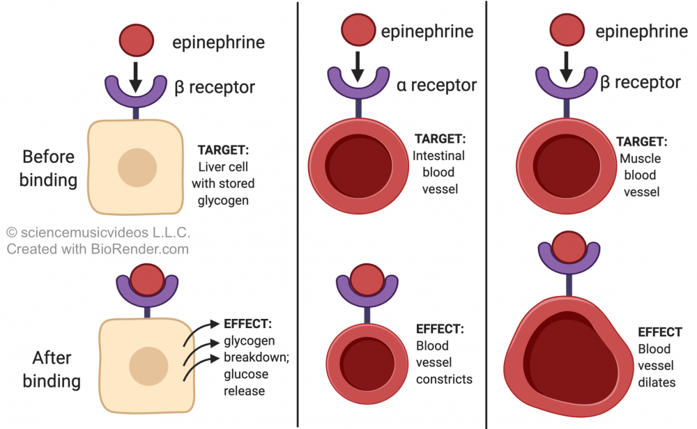
- Through their receptors, cells are “tuned” to respond to specific ligands (in much the same way that a radio receiver will pick up a specific station’s signal only when it’s tuned to the right frequency). However, a hormone might have a variety of targets throughout the body, and these targets, depending on the tissues that they’re a part of and their cells’ internal structure and chemistry, might respond to the hormone in different ways. For example, epinephrine has a variety of effects throughout the body.
-
- In the liver (as we’ve seen), epinephrine induces liver cells to break down glycogen into glucose
- In the smooth muscle tissue that lines the arteries that supply the intestines and the rest of the digestive system with blood, epinephrine induces muscle contraction. This reduces the diameter of these arteries and slows down the flow of blood to the digestive tract, which frees the body to divert blood away from the digestive system and towards the muscles of the skeletal system. This is also a part of the fight-or-flight response. From a survival perspective, the priority has to be getting away from that predator. You can digest your lunch when you’re safe (or, if you’ve vomited out your lunch because of fear, you can find more food to eat at a later time).
- In the smooth muscle surrounding the arteries that supply the muscles with blood, epinephrine causes muscle relaxation. This opens up these blood vessels, increasing the amount of blood that flows to the muscles. This is a useful adaptation if you’re escaping from or fighting off a predator.
SUMMARY: one hormone, different effects. We’ll see this again in our study of steroid hormones in the next tutorial.
What’s next?
- Proceed to Topics 4.2 – 4.3, part 4: Hormones and Gene Expression (the next tutorial in AP Bio Unit 4).

