1. Introduction
In response to the binding of a ligand with a membrane receptor, there are many mechanisms by which cells change their internal activities. In what follows, we’re going to focus on only one: G-protein coupled receptors. That’s because these are the receptors found in the glycogen-storing liver cells that respond to epinephrine by converting their glycogen into glucose (which is the story we’ve been trying to follow). Let’s see how.
2. G-protein Coupled Receptors
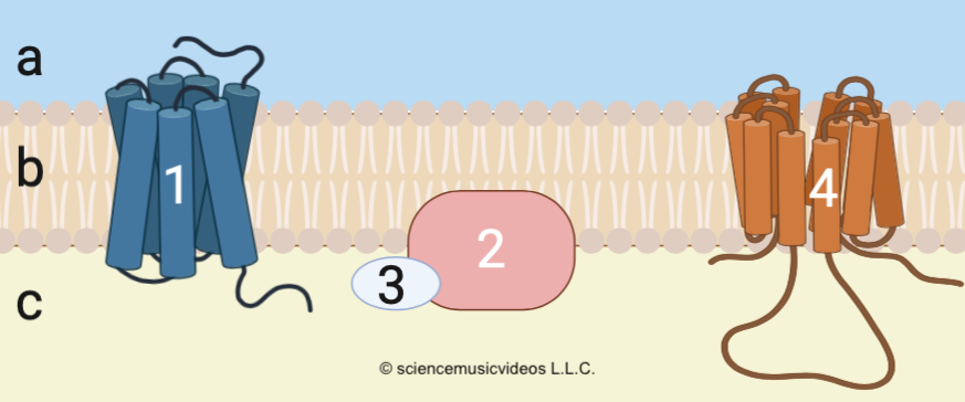
A G-protein coupled receptor is exactly what it sounds like. It’s a membrane receptor (“1,” at left) that works with a nearby membrane-embedded protein called a G-protein (“2”). The name “G protein” relates to the interaction between these proteins and either GTP or GDP, two energy-transfer molecules that function very similarly to ATP and ADP. When a ligand (like epinephrine) binds with the receptor, the receptor activates the G-protein, which, in turn, activates a nearby membrane-embedded enzyme (“4”), which sends a signal into the cytoplasm.
Using the diagrams below, we’ll see how this works. Note that the Roman numerals below represent a time sequence.
- “I” is the same as what’s shown above. It represents the moment before epinephrine binds.
- “II” shows what happens at the moment of binding.
- “III” shows both the changes that happen in response to epinephrine binding and epinephrine’s detachment from the membrane receptor.
- “IV” shows the deactivation of the cell’s initial response to epinephrine.
Read below, looking at the diagram to follow the action. Note that you can scroll the text below the diagram while continuing to be able to look at the diagram.
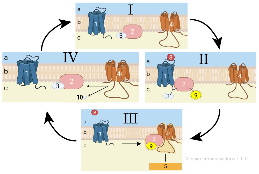
- Diagram “I” shows what the membrane (“b”), extracellular fluid (“a”), and cytoplasm (“c”) are like before epinephrine arrives at the cell. Embedded in the membrane is a receptor (“1”). This receptor is a transmembrane protein. Nearby, embedded in the membrane, is a G protein (represented by “2”). At this moment, before epinephrine binds, the G protein is inactive and bound to a molecule of GDP (at “3”). GDP stands for guanosine diphosphate. You can think of GDP exactly as you would think of ADP, the low energy counterpart of ATP. Finally, there’s a membrane-embedded enzyme (at “4”). This enzyme is currently inactive; activating it is a key step in bringing about a cellular response.
- In diagram “II,” epinephrine (“8”) binds with the receptor (“1”). Epinephrine’s binding at the extracellular side of the receptor changes the receptor’s shape and binding properties on its cytoplasmic side. This is analogous to the allosteric regulation of an enzyme, in which the binding of a regulator at an allosteric binding site changes the enzyme’s active site (and we’ll explain this in more detail immediately below). This change in the receptor’s cytoplasmic side enables it to bind with the G protein. This binding, in turn, changes the G protein so that it discharges its GDP, and binds instead with GTP (the high-energy form of this molecular duo).
- In diagram “III,” two things happen.
- First, the G protein, now bound to GTP, disconnects from the receptor and floats away within the membrane. Binding with GTP has changed the G protein’s shape so that it now can bind with the membrane-embedded enzyme shown at “4”. When the G protein binds with this enzyme it causes a conformational change in the enzyme (again, an allosteric shift), triggering the start of the cellular response. This response is indicated by “5,” and it’ll be the focus of the next tutorial.
- Second, epinephrine disconnects from the receptor. Note that the bonds between a ligand and a receptor are always weak and short-term, so nothing special needs to happen to get the ligand and receptor to disconnect.
- In step “IV,” GTP loses its terminal phosphate. You can see that phosphate group (at “10”) being released into the cytoplasm. This causes GTP to revert to GDP, and the G protein reverts to its inactive form. The G protein dissociates from the enzyme at “4,” which has the effect of deactivating the enzyme, which turns off this part of the cellular response.
The membrane is now ready to receive another signal, and whether or not that happens depends on whether the adrenal glands are releasing more epinephrine, which in turn depends on whether or not the initial stimulus for the fight-or-flight response is still present.
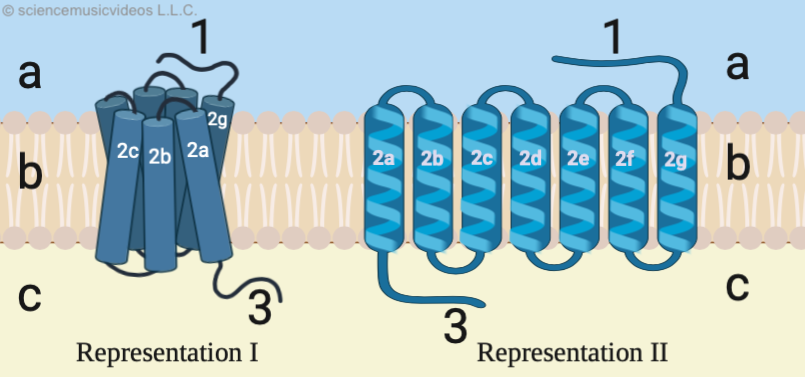 Finally, let’s hone in on the membrane receptor itself. The receptor is a tertiary-level protein. It consists of a ligand-binding site (shown at “1”), a series of seven alpha-helices that thread through the phospholipid bilayer, and a G-protein binding site (“3”). As stated above, you should think back to allosteric binding sites on enzymes to imagine the mechanism involved with a G-protein coupled receptor. When the ligand (epinephrine) binds with the ligand-binding site, it causes a conformational change that ripples through the entire receptor protein. Most importantly, it changes the shape of “3” so that this cytoplasmic portion of the receptor can bind with the G protein, setting off the cascade of reactions that we described above.
3. Quiz: Reception
[qwiz random = “true” style=”width: 750px !important; min-height: 400px !important;” qrecord_id=”sciencemusicvideosMeister1961-cell signaling: reception (v2.0)”]
[h] Quiz: Reception
[i]
[q json=”true” dataset_id=”SMV_Reception (cell communication)|63a6fc0cb8fff” question_number=”1″] Which number represents the signal binding site?
[textentry single_char=”true”]
[c]ID E=[Qq]
[f]IFllcy4g4oCcMeKAnSBpcyB0aGUgc2lnbmFsIGJpbmRpbmcgc2l0ZQ==[Qq]
[c]ICo=[Qq]
[f]IE5vLiBIZXJlJiM4MjE3O3MgYSBoaW50LiBUaGUgc2lnbmFsIGJpbmRpbmcgc2l0ZSBpcyBnb2luZyB0byBoYXZlIHRvIGZhY2UgdGhlIG91dHNpZGUgb2YgdGhlIGNlbGwgYW5kIGJlIHNvbWV0aGluZyB0aGF0IGEgc2lnbmFsIGNvdWxkIGJpbmQgd2l0aC4=
Cg==[Qq]
[q json=”true” dataset_id=”SMV_Reception (cell communication)|63a174707b7ff” question_number=”2″] Which number represents the receptor segment that interacts with the G-Protein?
[textentry single_char=”true”]
[c]ID M=[Qq]
[f]IFllcy4g4oCcM+KAnSByZXByZXNlbnRzIHRoZSBzZWdtZW50IG9mIHRoZSByZWNlcHRvciB0aGF0IGludGVyYWN0cyB3aXRoIGEgRy1wcm90ZWlu[Qq]
[c]ICo=[Qq]
[f]IE5vLiBUaGUgRy1wcm90ZWluIGlzIGVtYmVkZGVkIGluIHRoZSBpbm5lciBzaWRlIG9mIHRoZSBwbGFzbWEgbWVtYnJhbmUuIFdoaWNoIHBhcnQgb2YgdGhlIHJlY2VwdG9yIGNvdWxkIGludGVyYWN0IHdpdGggc29tZXRoaW5nIHRoYXQmIzgyMTc7cyBvbiB0aGUgaW5zaWRlIG9mIHRoZSBtZW1icmFuZT8=
Cg==[Qq]
[q json=”true” dataset_id=”SMV_Reception (cell communication)|639bc7937fbff” question_number=”3″] The alpha helices that embed the receptor in the membrane are represented by which number (type in a 1, 2, or a 3)?
[textentry single_char=”true”]
[c]ID I=[Qq]
[f]IFllcy4g4oCcMuKAnSByZXByZXNlbnRzIHRoZSBhbHBoYSBoZWxpY2VzIHRoYXQgZW1iZWQgdGhpcyByZWNlcHRvciBpbiB0aGUgY2VsbCBtZW1icmFuZS4=[Qq]
[c]ICo=[Qq]
[f]IE5vLiBIZXJlJiM4MjE3O3MgYSBoaW50OiBhIGhlbGl4IGlzIGxpa2UgYSBjb2lsZWQgc3ByaW5nLg==
Cg==[Qq]
[q json=”true” multiple_choice=”true” dataset_id=”SMV_Reception (cell communication)|6394f0b091fff” question_number=”4″] Which roman numeral shows the phase in which a ligand is binding with a receptor?
[c]IEk=[Qq]
[f]IE5vLiDigJxJ4oCdIHJlcHJlc2VudHMgdGhlIHBoYXNlIA==YmVmb3JlIHRoZSBsaWdhbmQgYmluZHMgd2l0aCB0aGUgcmVjZXB0b3Iu[Qq]
[c]IE lJ[Qq]
[f]IEV4Y2VsbGVudC4gJiM4MjIwO0lJJiM4MjIxOyByZXByZXNlbnRzIHRoZSBwaGFzZSB3aGVyZSBhIGxpZ2FuZCAoYSBob3Jtb25lIHN1Y2ggYXMgZXBpbmVwaHJpbmUsIGZvciBleGFtcGxlKSBpcyBiaW5kaW5nIHdpdGggdGhlIHJlY2VwdG9yLg==[Qq]
[c]IElJSQ==[Qq]
[f]IE5vLiAmIzgyMjA7SUlJJiM4MjIxOyBzaG93cyB0aGUgbGlnYW5kIGRpc3NvY2lhdGluZyBmcm9tIHRoZSByZWNlcHRvci4gV2hlcmUgZG8geW91IHNlZSB0aGUgbGlnYW5kIGJpbmRpbmcgd2l0aCB0aGUgcmVjZXB0b3I/[Qq]
[c]IElW[Qq]
[f]IE5vLiAmIzgyMjA7SVYmIzgyMjE7IHNob3dzIHRoZSBHIHByb3RlaW4gcmV2ZXJ0aW5nIHRvIGl0cyBpbmFjdGl2ZSBmb3JtIGFzIGl0IGNvbnZlcnRzIGl0cyBHVFAgdG8gR0RQLiBXaGVyZSBkbyB5b3Ugc2VlIHRoZSBsaWdhbmQgYmluZGluZyB3aXRoIHRoZSByZWNlcHRvcj8=
Cg==[Qq]
[q json=”true” multiple_choice=”true” dataset_id=”SMV_Reception (cell communication)|638e898fdefff” question_number=”5″] Which roman numeral shows the phase in which the G protein is becoming activated as it replaces its GDP with a GTP?
[c]IEk=[Qq]
[f]IE5vLiDigJxJ4oCdIHJlcHJlc2VudHMgdGhlIHBoYXNlIHdoZXJlIHRoZSBHIHByb3RlaW4gaXMgYm91bmQgdG8gR0RQIChtZWFuaW5nIHRoYXQgdGhlIEcgcHJvdGVpbiBpcyBpbiBhbiBpbmFjdGl2ZSBmb3JtKS4=[Qq]
[c]IE lJ[Qq]
[f]IEV4Y2VsbGVudC4gJiM4MjIwO0lJJiM4MjIxOyByZXByZXNlbnRzIHRoZSBwaGFzZSB3aGVyZSBhIGxpZ2FuZCAoYSBob3Jtb25lIHN1Y2ggYXMgZXBpbmVwaHJpbmUsIGZvciBleGFtcGxlKSBpcyBiaW5kaW5nIHdpdGggdGhlIHJlY2VwdG9yLiBUaGlzIGNoYW5nZXMgYSBzZWdtZW50IG9uIHRoZSByZWNlcHRvciwgd2hpY2ggaW4gdHVybiBtb2RpZmllcyB0aGUgRyBwcm90ZWluLCBjYXVzaW5nIGl0IHRvIHJlcGxhY2UgR0RQIHdpdGggR1RQLg==[Qq]
[c]IElJSQ==[Qq]
[f]IE5vLiAmIzgyMjA7SUlJJiM4MjIxOyBzaG93cyB0aGUgbGlnYW5kIGRpc3NvY2lhdGluZyBmcm9tIHRoZSByZWNlcHRvciBhbmQgYW4gYWN0aXZhdGVkIEcgcHJvdGVpbiBiaW5kaW5nIHdpdGggYSBtZW1icmFuZS1ib3VuZCBlbnp5bWUuIFdoaWNoIHBoYXNlIHNob3dzIHRoZSBhY3RpdmF0aW9uIG9mIHRoZSBHIHByb3RlaW4/[Qq]
[c]IElW[Qq]
[f]IE5vLiAmIzgyMjA7SVYmIzgyMjE7IHNob3dzIHRoZSBHIHByb3RlaW4gcmV2ZXJ0aW5nIHRvIGl0cyBpbmFjdGl2ZSBmb3JtIGFzIGl0IGNvbnZlcnRzIGl0cyBHVFAgdG8gR0RQLiBXaGljaCBwaGFzZSBzaG93cyB0aGUgYWN0aXZhdGlvbiBvZiB0aGUgRyBwcm90ZWluPw==
Cg==[Qq]
[q json=”true” multiple_choice=”true” dataset_id=”SMV_Reception (cell communication)|6385a92289bff” question_number=”6″] Which roman numeral shows the phase in which an activated G protein is activating a membrane-bound enzyme?
[c]IEk=[Qq]
[f]IE5vLiDigJxJ4oCdIHJlcHJlc2VudHMgdGhlIHBoYXNlIHdoZXJlIHRoZSBHIHByb3RlaW4gaXMgYm91bmQgdG8gR0RQIChtZWFuaW5nIHRoYXQgdGhlIEcgcHJvdGVpbiBpcyBpbiBhbiBpbmFjdGl2ZSBmb3JtKS4gV2hlcmUgZG8geW91IHNlZSBhbiBhY3RpdmF0ZWQgRyBwcm90ZWluIGludGVyYWN0aW5nIHdpdGggYSBtZW1icmFuZS1ib3VuZCBlbnp5bWU/[Qq]
[c]IElJ[Qq]
[f]IE5vLiAmIzgyMjA7SUkmIzgyMjE7IHJlcHJlc2VudHMgdGhlIHBoYXNlIHdoZXJlIGEgbGlnYW5kIChhIGhvcm1vbmUgc3VjaCBhcyBlcGluZXBocmluZSwgZm9yIGV4YW1wbGUpIGlzIGJpbmRpbmcgd2l0aCB0aGUgcmVjZXB0b3IuIFRoaXMgY2hhbmdlcyBhIHNlZ21lbnQgb24gdGhlIHJlY2VwdG9yLCB3aGljaCBpbiB0dXJuIG1vZGlmaWVzIHRoZSBHIHByb3RlaW4sIGNhdXNpbmcgaXQgdG8gcmVwbGFjZSBHRFAgd2l0aCBHVFAuIEJ1dCB0aGVyZSYjODIxNztzIG5vIGludGVyYWN0aW9uIGJldHdlZW4gdGhlIEcgcHJvdGVpbiBhbmQgdGhlIG1lbWJyYW5lLWJvdW5kIGVuenltZSAoYXQgJiM4MjIwOzQmIzgyMjE7KS4=[Qq]
[c]IElJ SQ==[Qq]
[f]IFllcy4gJiM4MjIwO0lJSSYjODIyMTsgc2hvd3MgdGhlIGxpZ2FuZCBkaXNzb2NpYXRpbmcgZnJvbSB0aGUgcmVjZXB0b3IgYW5kIGFuIGFjdGl2YXRlZCBHIHByb3RlaW4gYmluZGluZyB3aXRoIGEgbWVtYnJhbmUtYm91bmQgZW56eW1lLg==[Qq]
[c]IElW[Qq]
[f]IE5vLiAmIzgyMjA7SVYmIzgyMjE7IHNob3dzIHRoZSBHIHByb3RlaW4gcmV2ZXJ0aW5nIHRvIGl0cyBpbmFjdGl2ZSBmb3JtIGFzIGl0IGNvbnZlcnRzIGl0cyBHVFAgdG8gR0RQLiBXaGVyZSBkbyB5b3Ugc2VlIGFuIGFjdGl2YXRlZCBHIHByb3RlaW4gaW50ZXJhY3Rpbmcgd2l0aCBhIG1lbWJyYW5lLWJvdW5kIGVuenltZT8=
Cg==[Qq]
[q json=”true” multiple_choice=”true” dataset_id=”SMV_Reception (cell communication)|637fb1c4117ff” question_number=”7″] Which roman numeral shows the phase in which a G protein is reverting back to its inactive form, and in which cellular responses stop occurring.
[c]IEk=[Qq]
[f]IE5vLiDigJxJ4oCdIHJlcHJlc2VudHMgdGhlIHBoYXNlIHdoZXJlIHRoZSBHIHByb3RlaW4gaXMgYm91bmQgdG8gR0RQIChtZWFuaW5nIHRoYXQgdGhlIEcgcHJvdGVpbiBpcyBpbiBhbiBpbmFjdGl2ZSBmb3JtKS4gVGhpcyBxdWVzdGlvbiBpcyBhc2tpbmcgeW91IGhvdyB0aGUgRyBwcm90ZWluIGJlY2FtZSBkZWFjdGl2YXRlZCwgd2l0aCBhbiBhdHRhY2hlZCBHVFAgYmVjb21pbmcgY29udmVydGVkIHRvIEdEUC4=[Qq]
[c]IElJ[Qq]
[f]IE5vLiAmIzgyMjA7SUkmIzgyMjE7IHJlcHJlc2VudHMgdGhlIHBoYXNlIHdoZXJlIGEgbGlnYW5kIChhIGhvcm1vbmUgc3VjaCBhcyBlcGluZXBocmluZSwgZm9yIGV4YW1wbGUpIGlzIGJpbmRpbmcgd2l0aCB0aGUgcmVjZXB0b3IuIFRoaXMgY2hhbmdlcyBhIHNlZ21lbnQgb24gdGhlIHJlY2VwdG9yLCB3aGljaCBpbiB0dXJuIG1vZGlmaWVzIHRoZSBHIHByb3RlaW4sIGNhdXNpbmcgaXQgdG8gcmVwbGFjZSBHRFAgd2l0aCBHVFAuIFRoaXMgcXVlc3Rpb24gaXMgYXNraW5nIHlvdSBob3cgdGhlIEcgcHJvdGVpbiBiZWNvbWVzIGRlYWN0aXZhdGVkLCB3aXRoIGl0cyBhdHRhY2hlZCBHVFAgYmVpbmcgY29udmVydGVkIHRvIEdEUC4=[Qq]
[c]IElJSQ==[Qq]
[f]IE5vLiAmIzgyMjA7SUlJJiM4MjIxOyBzaG93cyB0aGUgbGlnYW5kIGRpc3NvY2lhdGluZyBmcm9tIHRoZSByZWNlcHRvciBhbmQgYW4gYWN0aXZhdGVkIEcgcHJvdGVpbiBiaW5kaW5nIHdpdGggYSBtZW1icmFuZS1ib3VuZCBlbnp5bWUuIFRoaXMgcXVlc3Rpb24gaXMgYXNraW5nIHlvdSBob3cgdGhlIEcgcHJvdGVpbiBiZWNvbWVzIA==ZGVhY3RpdmF0ZWQ=LCB3aXRoIGl0cyBhdHRhY2hlZCBHVFAgYmVpbmcgY29udmVydGVkIHRvIEdEUC4=[Qq]
[c]IE lW[Qq]
[f]IFllcy4gJiM4MjIwO0lWJiM4MjIxOyBzaG93cyB0aGUgRyBwcm90ZWluIHJldmVydGluZyB0byBpdHMgaW5hY3RpdmUgZm9ybSBhcyBpdCBjb252ZXJ0cyBpdHMgR1RQIHRvIEdEUC4=
Cg==[Qq]
[q json=”true” dataset_id=”SMV_Reception (cell communication)|6379dfa6577ff” question_number=”8″] In the series of diagrams below, which letter represents the cell membrane?
[textentry single_char=”true”]
[c]IG I=[Qq]
[f]IFllcy4gTGV0dGVy4oCcYuKAnSByZXByZXNlbnRzIHRoZSBjZWxsIG1lbWJyYW5l[Qq]
[c]ICo=[Qq]
[f]IE5vLiBIZXJlJiM4MjE3O3MgYSBoaW50LiBJZiAmIzgyMjA7YyYjODIyMTsgaXMgdGhlIGN5dG9wbGFzbSwgd2hhdCBsZXR0ZXIgaGFzIHRvIGJlIHRoZSBtZW1icmFuZT8=
Cg==[Qq]
[q json=”true” dataset_id=”SMV_Reception (cell communication)|63740d889d7ff” question_number=”9″] In the series of diagrams below, which number represents the receptor?
[textentry single_char=”true”]
[c]ID E=[Qq]
[f]IFllcy4g4oCcMeKAnSByZXByZXNlbnRzIHRoZSByZWNlcHRvci4=[Qq]
[c]ICo=[Qq]
[f]IE5vLiBIZXJlJiM4MjE3O3MgYSBoaW50LiBJZiAmIzgyMjA7OCYjODIyMTsgcmVwcmVzZW50cyB0aGUgbGlnYW5kICh0aGUgbW9sZWN1bGUgdGhhdCBiaW5kcyB3aXRoIHRoZSByZWNlcHRvciksIHRoZW4gd2hpY2ggbnVtYmVyIGhhcyB0byByZXByZXNlbnQgdGhlIHJlY2VwdG9yIGl0c2VsZj8=
Cg==[Qq]
[q json=”true” dataset_id=”SMV_Reception (cell communication)|636e3b6ae37ff” question_number=”10″] In the series of diagrams below, which number represents the G protein?
[textentry single_char=”true”]
[c]ID I=[Qq]
[f]IFllcy4g4oCcMuKAnSByZXByZXNlbnRzIHRoZSBHIHByb3RlaW4u[Qq]
[c]ICo=[Qq]
[f]IE5vLiBIZXJlJiM4MjE3O3MgYSBoaW50LiBUaGUgRyBwcm90ZWluIGlzIGFjdGl2YXRlZCBieSB0aGUgcmVjZXB0b3IgYW5kIHRoZW4gZ29lcyBvbiB0byBhY3RpdmF0ZSBhIG1lbWJyYW5lLWJvdW5kIGVuenltZSAoc3VjaCBhcyB0aGUgb25lIGRlcGljdGVkIGluIG51bWJlciAmIzgyMjA7NCYjODIyMTspLiBPZiBhbGwgb2YgdGhlIGNvbXBvbmVudHMgb2YgdGhpcyBzeXN0ZW0sIHdoaWNoIG9uZSBjb3VsZCBwbGF5IHN1Y2ggYSByb2xlPw==
Cg==[Qq]
[q json=”true” dataset_id=”SMV_Reception (cell communication)|6367f98aeebff” question_number=”11″] In the series of diagrams below, which number represents GDP?
[textentry single_char=”true”]
[c]ID M=[Qq]
[f]IFllcy4g4oCcM+KAnSByZXByZXNlbnRzIEdEUCAoZ3Vhbm9zaW5lIGRpcGhvc3BoYXRlKS4gR0RQIGlzIGFuYWxvZ291cyB0byBBRFAgKGFkZW5vc2luZSBkaXBob3NwaGF0ZSkgaW4gdGhhdCBpdCBjYW4gYmUgcGhvc3Bob3J5bGF0ZWQgdG8gYmVjb21lIHRoZSBoaWdoZXIgZW5lcmd5LCBhY3RpdmF0ZWQgZm9ybTogR1RQIG9yIGd1YW5vc2luZSB0cmlwaG9zcGhhdGUuIFRoYXQmIzgyMTc7cyBleGFjdGx5IHdoYXQgaGFwcGVucyBpbiB0aGlzIHN5c3RlbTogQXMgR0RQIGJlY29tZXMgR1RQLCB0aGUgRyBwcm90ZWluIGJlY29tZXMgYWN0aXZhdGVkLCBlbmFibGluZyBpdCB0byBpbnRlcmFjdCB3aXRoIGEgbWVtYnJhbmUtYm91bmQgZW56eW1lIHRvIGJyaW5nIGFib3V0IHRoZSBjZWxsdWxhciByZXNwb25zZSB0byB0aGUgYmluZGluZyBvZiB0aGUgbGlnYW5kLg==[Qq]
[c]ICo=[Qq]
[f]IE5vLiBIZXJlJiM4MjE3O3MgYSBoaW50LiBHRFAgKChndWFub3NpbmUgZGlwaG9zcGhhdGUpIGlzIGEgbG93LWVuZXJneSBmb3JtIG9mIEdUUCAodHJpcGhvc3BoYXRlKS4gV2hlbiBHRFAgaXMgYm91bmQgdG8gYSBHIHByb3RlaW4sIHRoYXQgcHJvdGVpbiBpcyBlc3NlbnRpYWxseSBpbmVydC4gV2hhdCBkbyB5b3Ugc2VlIHRoYXQmIzgyMTc7cyBib3VuZCB0byB0aGUgRyBwcm90ZWluIHRoYXQgY2FuIGJlIHRyYW5zZm9ybWVkIGZyb20gYSBsb3cgZW5lcmd5IHN0YXRlIGludG8gYSBoaWdoIGVuZXJneSBzdGF0ZT8=
Cg==[Qq]
[q json=”true” dataset_id=”SMV_Reception (cell communication)|636271eeb13ff” question_number=”12″] In the series of diagrams below, which number represents a membrane-bound enzyme which, when activated, can initiate a cellular response?
[textentry single_char=”true”]
[c]ID Q=[Qq]
[f]IFllcy4g4oCcNOKAnSByZXByZXNlbnRzIGEgbWVtYnJhbmUtYm91bmQgZW56eW1lLiBXaGVuIHRoaXMgZW56eW1lIGJlY29tZXMgYWN0aXZhdGVkLCBpdCBjYW4sIGluIHR1cm4sIGFjdGl2YXRlIG90aGVyIG1vbGVjdWxlcyB3aGljaCBjYW4gYnJpbmcgYWJvdXQgYSBjZWxsdWxhciByZXNwb25zZS4=[Qq]
[c]ICo=[Qq]
[f]IE5vLiBIZXJlJiM4MjE3O3MgYSBoaW50LiBMb29rIGZvciBzb21ldGhpbmcgdGhhdCYjODIxNztzIGJvdW5kIHRvIHRoZSBtZW1icmFuZSB0aGF0JiM4MjE3O3Mgbm90IGEgcmVjZXB0b3Igb3IgYSBHIHByb3RlaW4u
Cg==[Qq]
[q json=”true” dataset_id=”SMV_Reception (cell communication)|635c9fd0f73ff” question_number=”13″] In the series of diagrams below, which number represents GTP?
[textentry single_char=”true”]
[c]ID k=[Qq]
[f]IFllcy4g4oCcOeKAnSByZXByZXNlbnRzIEdUUC4gV2hlbiBHVFAgYmluZHMgdG8gdGhlIEcgcHJvdGVpbiwgdGhlIEcgcHJvdGVpbiBiZWNvbWVzIGFjdGl2YXRlZCwgYWxsb3dpbmcgaXQgdG8gaW50ZXJhY3Qgd2l0aCBtZW1icmFuZS1ib3VuZCBlbnp5bWVzIHRoYXQgY2FuLCBpbiB0dXJuLCBpbml0aWF0ZSB0aGUgY2VsbHVsYXIgcmVzcG9uc2Uu[Qq]
[c]ICo=[Qq]
[f]IE5vLiBIZXJlJiM4MjE3O3MgYSBoaW50LiBHVFAgaXMgYW4gYWN0aXZhdGVkIGZvcm0gb2YgR0RQLiBJbiB0aGlzIHN5c3RlbSwgYm90aCBjYW4gYmUgZm91bmQgYm91bmQgdG8gdGhlIEcgcHJvdGVpbi4gU28sIGxvb2sgZm9yIHNvbWV0aGluZyB0aGF0JiM4MjE3O3MgYm91bmQgdG8gYSBHIHByb3RlaW4sIGFuZCB3aGljaCBpcyByZXByZXNlbnRlZCBhcyBiZWluZyBpbiBhIGhpZ2gtZW5lcmd5IHN0YXRlLg==
Cg==[Qq]
[q json=”true” dataset_id=”SMV_Reception (cell communication)|63565df1027ff” question_number=”14″] In the series of diagrams below, which number represents the cytoplasmic response to the binding of a ligand?
[textentry single_char=”true”]
[c]ID U=[Qq]
[f]IFllcy4g4oCcNeKAnSByZXByZXNlbnRzIHRoZSBjeXRvcGxhc21pYyByZXNwb25zZSB0byB0aGUgYmluZGluZyBvZiBhIGxpZ2FuZC4=[Qq]
[c]ICo=[Qq]
[f]IE5vLiBUaGUgY3l0b3BsYXNtaWMgcmVzcG9uc2UgY29tZXMgYWJvdXQgYXMgYSByZXN1bHQgb2YgYWN0aXZpdGllcyBieSBtZW1icmFuZS1ib3VuZCBlbnp5bWVzIHRoYXQgaGF2ZSBiZWVuIGFjdGl2YXRlZCBieSB0aGUgRyBwcm90ZWluICh3aGljaCB3YXMgYWN0aXZhdGVkIGJ5IHRoZSByZWNlcHRvciwgd2hpY2ggd2FzIGFjdGl2YXRlZCBieSB0aGUgbGlnYW5kKS4gU3R1ZHkgdGhlIGRpYWdyYW0sIGFuZCBmaW5kIHRoZSBjeXRvcGxhc21pYyByZXNwb25zZSB0aGF0JiM4MjE3O3MgYXQgdGhlIGVuZCBvZiB0aGlzIGNoYWluLg==
Cg==[Qq]
[q json=”true” multiple_choice=”true” dataset_id=”SMV_Reception (cell communication)|634fd18f913ff” question_number=”15″] The G protein can be deactivated, bringing the cellular response to a close, when the GTP that’s bound to the G protein is converted to GDP. In the series of diagrams below, which number represents the phosphate group that is released during this conversion?
[c]IDI=[Qq]
[f]IE5vLiDigJwy4oCdIHJlcHJlc2VudHMgdGhlIEcgcHJvdGVpbi4=[Qq]
[c]IDM=[Qq]
[f]IE5vLiDigJwz4oCdIHJlcHJlc2VudHMgdGhlIEdEUC4=[Qq]
[c]IDk=[Qq]
[f]IE5vLiDigJw54oCdIHJlcHJlc2VudHMgdGhlIEdUUC4=[Qq]
[c]ID Ew[Qq]
[f]IFdheSB0byBnby4g4oCcMTDigJ0gcmVwcmVzZW50cyB0aGUgcGhvc3BoYXRlIHRoYXQmIzgyMTc7cyByZW1vdmVkIGZyb20gR1RQLiBMb3NzIG9mIHRoaXMgcGhvc3BoYXRlIGNvbnZlcnRzIEdUUCB0byBHRFAsIHdoaWNoIHJlbmRlcnMgdGhlIEcgcHJvdGVpbiBpbnRvIGFuIGluYWN0aXZlIGZvcm0u[Qq]
[q json=”true” dataset_id=”SMV_Reception (cell communication)|634a49f353bff” question_number=”16″] In the diagram below, number 3 is [hangman]
[c]IEdEUA==[Qq]
[f]IEdvb2Qh[Qq]
[q json=”true” dataset_id=”SMV_Reception (cell communication)|6344e797d47ff” question_number=”17″] In the diagram below, number 9 is [hangman]
[c]IEdUUA==[Qq]
[f]IEdvb2Qh[Qq]
[q json=”true” dataset_id=”SMV_Reception (cell communication)|633f157a1a7ff” question_number=”18″] In the diagram below, number 4 is a membrane embedded [hangman]
[c]IGVuenltZQ==[Qq]
[f]IEdvb2Qh[Qq]
[q json=”true” dataset_id=”SMV_Reception (cell communication)|6339689d1ebff” question_number=”19″] If the diagram below were about mobilization of glucose from glycogen storing liver cells, then number 8 would be the hormone [hangman]
[c]IGVwaW5lcGhyaW5l[Qq]
[f]IEdvb2Qh[Qq]
[q json=”true” dataset_id=”SMV_Reception (cell communication)|6333bbc022fff” question_number=”20″] In the diagram below, number 10 is a [hangman] group.
[c]IHBob3NwaGF0ZQ==[Qq]
[f]IEdvb2Qh[Qq]
[q json=”true” dataset_id=”SMV_Reception (cell communication)|632dc461aabff” question_number=”21″] In the diagram below, number 1 is a G-coupled protein [hangman].
[c]IHJlY2VwdG9y[Qq]
[f]IEdvb2Qh[Qq]
[x][restart]
[/qwiz]
What’s next?
- Proceed to the next tutorial in AP Bio Unit 4: Topics 4.2 – 4.3, Part 3: Signal Transduction