Click the following link for a DNA Structure and Replication Student Learning Guide (coming soon)
1. Introduction
In the last tutorial, we looked at the structure of DNA.

DNA’s structure lends itself to replication. That’s essential for a molecule that gets passed from one generation to the next during reproduction, and from one cell to the next as an organism grows and develops.
2. Replication means “copying DNA”
DNA replication is one of life’s most essential processes. In your body, it’s happening all the time. Every time one of your cells divides—a key part of how you grow and maintain yourself — all of the DNA in that cell needs to be replicated. On a very big-picture level, here’s what that looks like. Note how the chromosomes in the parent cell on the left are being copied (arrow 1) then pulled apart (arrows 2 and 3) to create daughter cells (arrow 4) that have chromosomes that are exact duplicates of those in the parent cell.
Here’s what 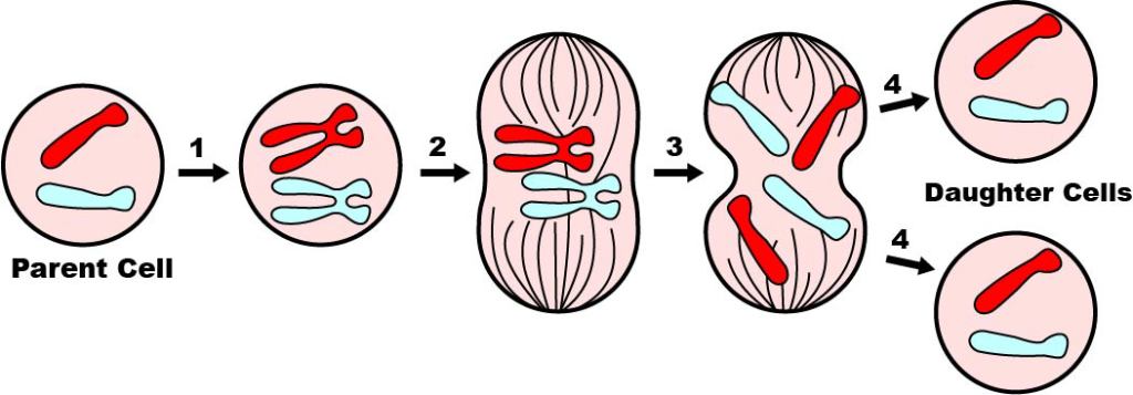
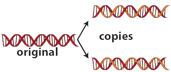
If you go down to a molecular level, here’s what replication looks like. Two new molecules of “daughter” DNA are made from one molecule of “parent” DNA.
DNA’s structure explains how cells can do this. Here’s how I explain this in the first two verses of my DNA Replication music video.
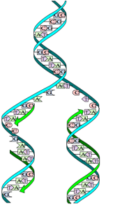

DNA’s structure with its bases complementary
Makes replication easy, but not quite elementary
Since A only bonds with T and C with G
The Double Helix seems to copy naturally,
Or as Crick and Watson said:
“It has not escaped our notice that the specific pairing
We have postulated immediately suggests
A possible copying mechanism
For the genetic material”
The last verse is a quote from a paper written in 1953 that first correctly described the structure of DNA. The paper was written by James Watson and Francis Crick, who were the co-discoverers of the structure of DNA. Their work relied heavily on the work of two other scientists:, Rosalind Franklin and Maurice Wilkins.
In what follows, we’ll explain how replication works.
3. During DNA Replication, each strand serves as a template for the synthesis of a complementary strand
Here’s a big picture view of what happens during DNA Replication.
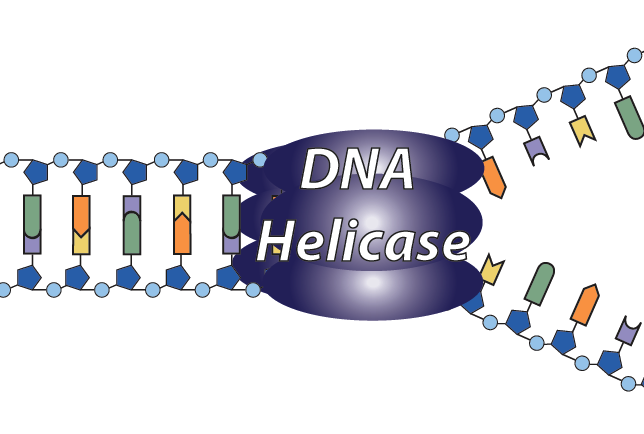
- An enzyme separates the single strands that make up the DNA double helix. The enzyme that does this is called helicase. That’s easy to remember because it’s the enzyme that opens the helix. Helicase does this by breaking the hydrogen bonds that connect the base pairs.
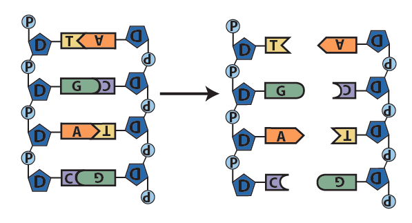
Step 1: The enzyme helicase (not shown) breaks the hydrogen bonds holding the two strands of the double helix together. This separates the DNA into into two single strands. - With the nitrogenous bases (A, T, C, or G) on each strand exposed, each single strand now serves as a template for synthesis of a new strand. A template is “a molecule … that serves as a pattern for the generation of another molecule” (source: Merriam-Webster on-line dictionary). For example, consider the single strand on the left side below. The base “T” is in the first position. Because of the base pairing rules, the only nucleotide that could bond with “T” is “A.” Similarly, the only nucleotide that could bond with the second nucleotide, “G,” is “C.” The key idea is that the information in one strand gives the cell the information needed to build the complementary strand.
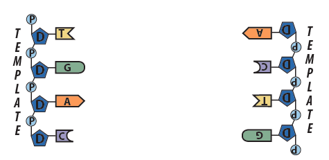
Step 2: Each strand serves as a template for synthesis of a new strand - Following the base pairing rules, complementary nucleotides form hydrogen bonds with the nucleotides on the template strands. Where do these new nucleotides come from? It depends. Plant cells can synthesize the nucleotides from scratch. Animals synthesize them from other molecules that they’ve eaten,
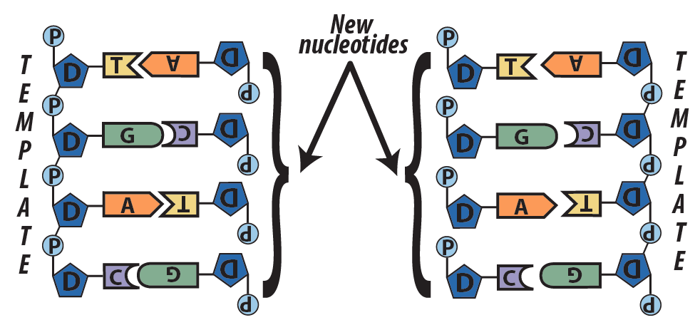
Step 3: new complementary nucleotides form hydrogen bonds with the nitrogenous basis in the parent strands. - Another enzyme called DNA polymerase creates covalent bonds between the sugars and the phosphates of adjacent nucleotides on the newly synthesized strand. Once these sugar phosphate bonds have been created, DNA replication is complete. Where there was one DNA molecule, now there are two.
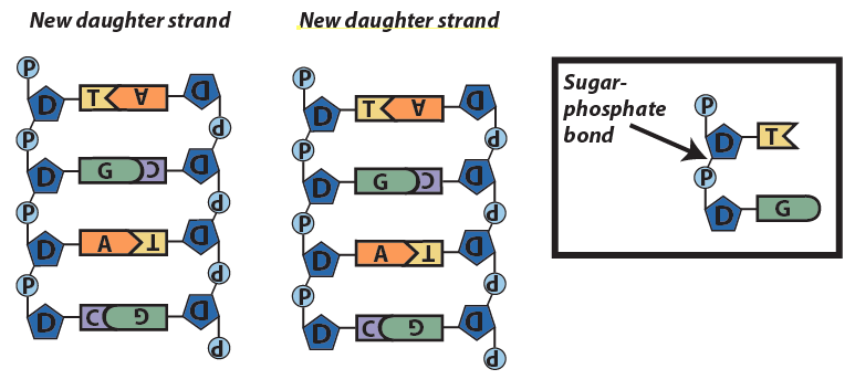
Step 4: Enzymes seal the new strands together with sugar-phosphate bonds.
This model of replication is called semi-conservative: in each new daughter molecule, one strand is old (from the parent), and one strand is new. You can see this in the color coding of the diagram below. The original DNA is colored red. The newly synthesized strands are colored orange.

Here’s the same explanation in verse:
You first unzip the DNA in one or more places,
Breaking hydrogen bonds to separate the bases.
Each resulting single strand serves as a template,
Allowing enzymes to replicate
New strands with complementary bases that match
And through hydrogen bonds these bases attach
Each nucleotide now bonds to the next
Through a sugar-phosphate bond they connect
Meselsohn and Stahl proved in ‘58 (read about their experiment)
That this is how the double helix replicates
One strand new, the parent strand preserved,
In other words the whole thing is semi-conserved
4. Checking Understanding: DNA Replication (the big picture)
[qwiz random = “false” qrecord_id=”sciencemusicvideosMeister1961-DNA Replication, Big Picture (HS)”]
[h] DNA replication: The Big Picture
[i]
[q] Copying of DNA is also know known as [hangman]
[c]IHJlcGxpY2F0aW9u[Qq]
[f]IEdvb2Qh[Qq]
[q] During replication, each strand acts as a [hangman] for synthesis of a new strand.
[c]IHRlbXBsYXRl[Qq]
[f]IEdvb2Qh[Qq]
[q] To start the process of replication, an enzyme called helicase needs to break the [hangman] bonds (shown below) that hold the base pairs together.
[c]IGh5ZHJvZ2Vu[Qq]
[f]IEdvb2Qh[Qq]
[q] Replication ends as enzymes create covalent bonds between the sugar [hangman] on one nucleotide and the phosphate group on the next nucleotides.
[c]IGRlb3h5cmlib3Nl[Qq]
[f]IEdyZWF0IQ==[Qq]
[q] Replication ends as DNA polymerase creates a covalent bonds between the deoxyribose sugar on one nucleotide and a [hangman] group on the next nucleotide.
[c]IHBob3NwaGF0ZQ==[Qq]
[f]IEZhbnRhc3RpYyE=[Qq]
[q] The result of replication is two daughter DNA molecules, each of which has one [hangman] strand and one old strand.
[c]IG5ldw==[Qq]
[f]IENvcnJlY3Qh[Qq]
[q] Because [hangman] results in daughter strands that are half new and half old, replication is called semi-conservative.
[c]IHJlcGxpY2F0aW9u[Qq]
[f]IEdyZWF0IQ==[Qq]
[q] DNA replication works because the nucleotide “A” is [hangman] to the nucleotide “T” (and the same is true of “C” and “G”)
[c]IGNvbXBsZW1lbnRhcnk=[Qq]
[f]IENvcnJlY3Qh[Qq]
[q multiple_choice=”true”] At the start of replication, an enzyme breaks open the helix. That enzyme is called
[c]IGhlbG ljYXNl[Qq]
[f]IEV4Y2VsbGVudCEgVGhlIGVuenltZSB0aGF0IGJyZWFrcyBvcGVuIHRoZSBoZWxpeCBpcyBjYWxsZWQgaGVsaWNhc2U=[Qq]
[c]cG9seW1lcmFzZQ==[Qq]
[f]IE5vLiBETkEgcG9seW1lcmFzZSBjcmVhdGVzIHN1Z2FyLXBob3NwaGF0ZSBib25kcyBiZXR3ZWVuIG9uZSBudWNsZW90aWRlIGFuZCB0aGUgbmV4dC4=[Qq]
[q multiple_choice=”true”] During replication, the enzyme breaks that creates sugar-phosphate bonds between one nucleotide and the next is
[c]IGhlbGljYXNl[Qq]
[f]IE5vLiBIZWxpY2FzZSBicmVha3Mgb3BlbiB0aGUgZG91YmxlIGhlbGl4IGJ5IGJyZWFraW5nIGh5ZHJvZ2VuIGJvbmRzIGJldHdlZW4gYmFzZSBwYWlycy4=[Qq]
[c]IEROQSBwb2 x5bWVyYXNl[Qq]
[f]IEV4Y2VsbGVudC4gRE5BIHBvbHltZXJhc2UgY3JlYXRlcyBzdWdhci1waG9zcGhhdGUgYm9uZHMgYmV0d2VlbiBvbmUgbnVjbGVvdGlkZSBhbmQgdGhlIG5leHQu[Qq]
[/qwiz]
5. How DNA replication occurs in cells
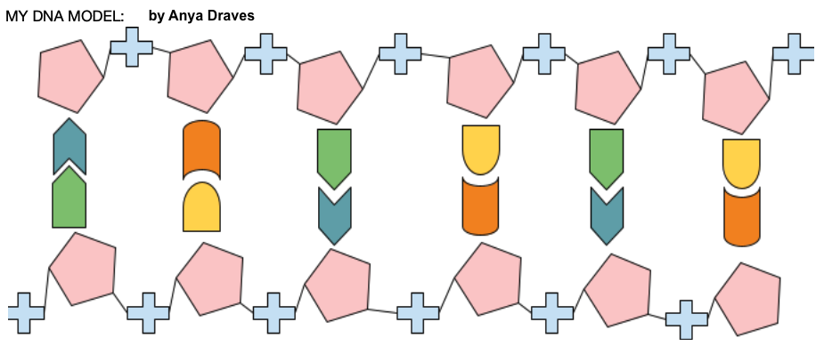
Imagine that you were given the task of taking a paper cutout model of DNA (like the one at left) and replicating it. You’d probably find this to be pretty easy. After all, you can see the bases, think of the base pairing rules, and manipulate the model (pulling it apart, adding new nucleotides, etc.) to create two new daughter strands from the parent strand.
How can cells, which can’t see or think, perform this task?
Cells do this all by enzymes, which have to do everything by touch and feel. You learned about two of these enzymes above. What follows expands and deepens that discussion.
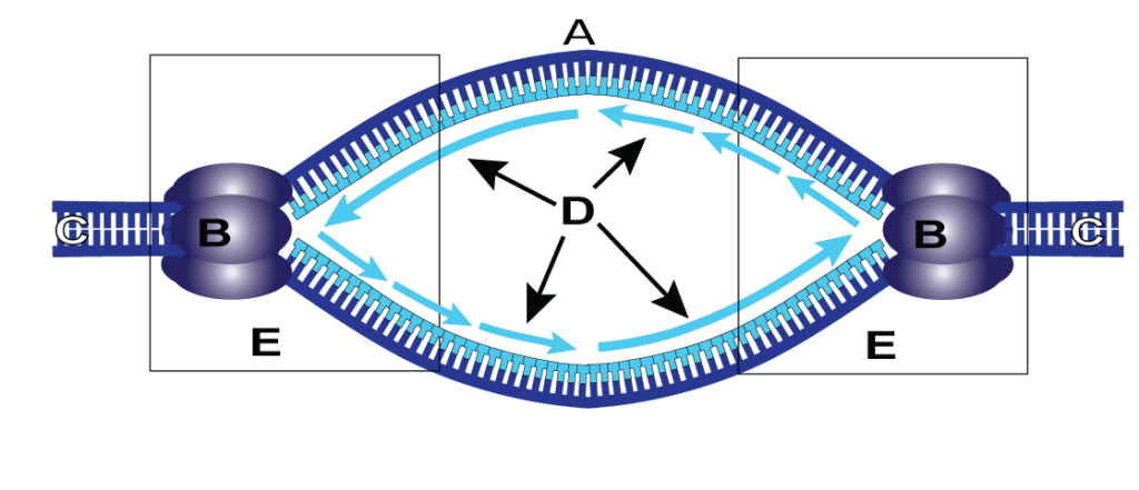
DNA helicase (represented by “B” in the diagram below) starts the process. Helicase finds a sequence of bases called the origin of replication (A). The origin of replication acts as a signal that says “start replicating here.” Once it finds the origin of replication, DNA helicase breaks open the double helix by breaking the hydrogen bonds between complementary bases.

The structure that results from the opening of the helix is called a replication fork.
Replication forks are always created in pairs, creating a replication bubble (the entire structure below). In the diagram below, the original is at shown in dark blue at C. Helicases are shown at B. Newly synthesized DNA is shown in light blue at “D.” The two replication forks are at “E,” and the origin of replication is at “A.”
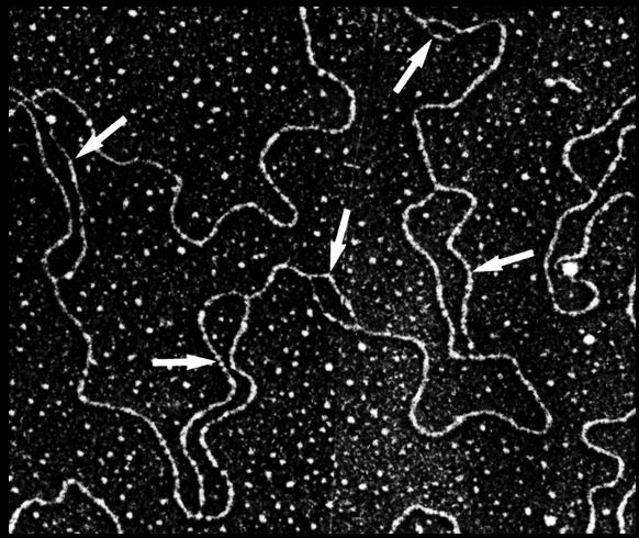
 Check out this amazing electron micrograph. It’s showing DNA replication in the nucleus of a eukaryotic cell. Each of the white arrows is pointing to a replication bubble.
With the nitrogenous bases of the template strand exposed, the cell can now create a complementary DNA strand. As we learned above, the enzyme that does this is DNA polymerase (“G” below). DNA polymerase rides along the template strand. As new nucleotides form hydrogen bonds with their complements on the template strand, DNA polymerase catalyzes a sugar-phosphate bond between the new nucleotide and the previous one. 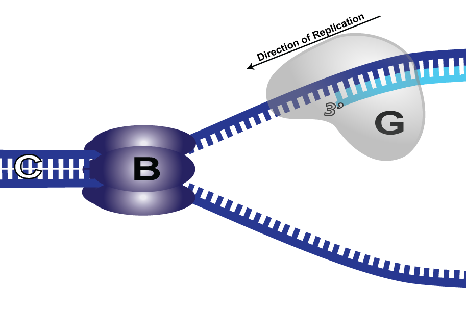
6. Here’s What it Really Looks Like…
The process I described above is simplified model of some of the key events of DNA replication. But it’s actually much more complex. Watch this computer animation from the Howard Hughes Medical Institute. Unbelievably, the model below is also a simplified version of the process.
7. Prokaryotic v. Eukaryotic DNA Replication
Because of the different sizes and organization of the genomes of prokaryotic and eukaryotic organisms, there are differences in how DNA replication proceeds in these two groups of organisms.
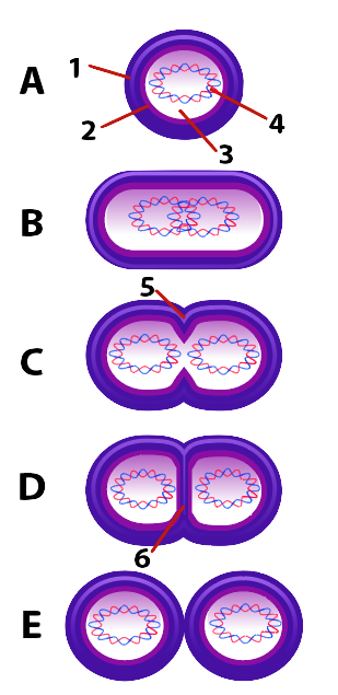
We’ll start with DNA replication in bacteria. In the diagram below, “A” represents the parent cell, and 4 represents its DNA. The other parts shown are the cytoplasm at “3,” the membrane at “4,” and an outer capsule (found in many bacteria, at “1”).
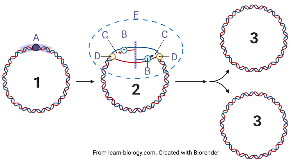
Bacteria have a single, circular (or looped) chromosome (as shown in “1” below). Replication begins at an origin of replication, shown at “A” in diagram 1. Helicases (at “D”) open up the helix, creating two replication forks (at “C”). DNA polymerases (at “B”) synthesize new strands of DNA. “E” represents the ever-widening replication bubble.
When replication is complete we have two daughter strand, each a perfect copy of the parent strand at “1.”
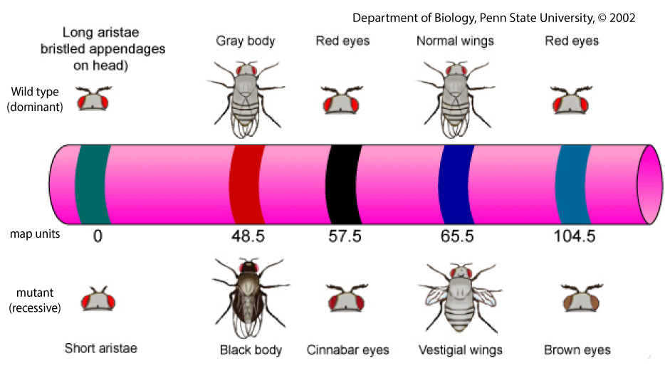
In terms of DNA replication, eukaryotic DNA differs from prokaryotic way in two notable ways. First, eukaryotic DNA is organized into linear chromosomes. That means that rather than a circular loop of DNA, the chromosomes have a beginning and an end. The diagram below shows a chromosome in a fruit fly. Fruit flies are among the most studied organisms on Earth. This diagram shows some of the genes on this chromosome, and the distance between these genes.
Because eukaryotic chromosomes are linear (and much longer than bacterial chromosomes), replication involves multiple replication bubbles (shown at E below). In each replication bubble, there will be helicases (D) and polymerases (B) working away.

8. Quiz: DNA Replication (with some DNA structure review)
That’s all you need to know about DNA replication. Take the quiz below to consolidate your learning.
[qwiz style=” width: 700px !important; ” qrecord_id=”sciencemusicvideosMeister1961-DNA Replication (HS)”]
[h] DNA replication in cells
[q]For “A”, just the first word. For “B,” it’s the name of the enzyme.
[c]IGhlbGljYXNl[Qq]
[f]IEV4Y2VsbGVudCE=[Qq]
[c]IG9yaWdpbg==[Qq]
[f]IEdyZWF0IQ==[Qq]
[q] In the diagram below, the origin of replication is at
[textentry single_char=”true”]
[c]IE E=[Qq]
[f]IEV4Y2VsbGVudC4gJiM4MjIwO0EmIzgyMjE7IGlzIHRoZSBvcmlnaW4gb2YgcmVwbGljYXRpb24u[Qq]
[c]IEVudGVyIHdvcmQ=[Qq]
[f]IE5vLg==[Qq]
[c]ICo=[Qq]
[f]IE5vLiBZb3UmIzgyMTc7cmUgbG9va2luZyBmb3IgdGhlIHNwb3Qgd2hlcmUgcmVwbGljYXRpb24gYmVnaW5zLg==[Qq]
[q] In the diagram below, DNA polymerase is at
[textentry single_char=”true”]
[c]IE I=[Qq]
[f]IE5pY2UuICYjODIyMDtCJiM4MjIxOyByZXByZXNlbnRzIEROQSBwb2x5bWVyYXNl[Qq]
[c]IEVudGVyIHdvcmQ=[Qq]
[f]IE5vLg==[Qq]
[c]ICo=[Qq]
[f]IE5vLiBZb3UmIzgyMTc7cmUgbG9va2luZyBmb3IgdGhlIGVuenltZSB0aGF0JiM4MjE3O3Mgc3ludGhlc2l6aW5nIG5ldyBETkEu[Qq]
[q] In the diagram below, helicase is at
[textentry single_char=”true”]
[c]IE Q=[Qq]
[f]IEV4Y2VsbGVudC4gJiM4MjIwO0QmIzgyMjE7IHJlcHJlc2VudHMgaGVsaWNhc2U=[Qq]
[c]IEVudGVyIHdvcmQ=[Qq]
[f]IE5vLg==[Qq]
[c]ICo=[Qq]
[f]IE5vLiBZb3UmIzgyMTc7cmUgbG9va2luZyBmb3IgdGhlIGVuenltZSB0aGF0IG9wZW5zIHVwIEROQS4=[Qq]
[q] In the diagram below, what letter represents a replication bubble?
[textentry single_char=”true”]
[c]IE U=[Qq]
[f]IE5pY2Ugam9iLiAmIzgyMjA7RCYjODIyMTsgcmVwcmVzZW50cyB0aGUgcmVwbGljYXRpb24gYnViYmxlLg==[Qq]
[c]IEVudGVyIHdvcmQ=[Qq]
[f]IFNvcnJ5LCB0aGF0JiM4MjE3O3Mgbm90IGNvcnJlY3Qu[Qq]
[c]ICo=[Qq]
[f]IE5vLiBZb3UmIzgyMTc7cmUgbG9va2luZyBmb3IgdGhlIHN0cnVjdHVyZSB0aGF0IGZvcm1zIGZyb20gdHdvIHJlcGxpY2F0aW9uIGZvcmtzLg==[Qq]
[q] In the diagram below, you know what’s being represented is DNA replication that would occur in a [hangman] cell. That’s because the DNA is [hangman].
[c]IGJhY3RlcmlhbA==[Qq]
[f]IEdyZWF0IQ==[Qq]
[c]IGNpcmN1bGFy[Qq]
[f]IEdyZWF0IQ==[Qq]
[q] In the diagram below, you know what’s being represented is DNA replication that would occur in a [hangman] cell. That’s because the DNA is [hangman].
[c]IGV1a2FyeW90aWM=[Qq]
[f]IEdvb2Qh[Qq]
[c]IGxpbmVhcg==[Qq]
[f]IENvcnJlY3Qh[Qq]
[q]
[c]IHBvbHltZXJhc2U=[Qq]
[f]IENvcnJlY3Qh[Qq]
[c]IGhlbGljYXNl[Qq]
[f]IEV4Y2VsbGVudCE=[Qq]
[q] In the diagram below, what letter represents the origin of replication?
[textentry single_char=”true”]
[c]IE E=[Qq]
[f]IEdvb2Qgd29yay4gJiM4MjIwO0EmIzgyMjE7IHJlcHJlc2VudHMgdGhlIG9yaWdpbiBvZiByZXBsaWNhdGlvbi4=[Qq]
[c]IEVudGVyIHdvcmQ=[Qq]
[f]IE5vLg==[Qq]
[c]ICo=[Qq]
[f]IE5vLiBZb3UmIzgyMTc7cmUgbG9va2luZyBmb3IgdGhlIHBsYWNlIHdoZXJlIHJlcGxpY2F0aW9uIHN0YXJ0cy4=[Qq]
[q] In the diagram below, what letter represents newly synthesized complementary DNA?
[textentry single_char=”true”]
[c]IE Q=[Qq]
[f]IE5pY2Ugam9iLiAmIzgyMjA7RCYjODIyMTsgcmVwcmVzZW50cyBuZXdseSBzeW50aGVzaXplZCBETkEu[Qq]
[c]IEVudGVyIHdvcmQ=[Qq]
[f]IE5vLCB0aGF0JiM4MjE3O3Mgbm90IGNvcnJlY3Qu[Qq]
[c]ICo=[Qq]
[f]IE5vLiBIZXJlJiM4MjE3O3MgYSBoaW50LiBJZiAmIzgyMjA7QyYjODIyMTsgcmVwcmVzZW50cyB0aGUgcGFyZW50IHN0cmFuZCwgd2hhdCBsZXR0ZXIgd291bGQgcmVwcmVzZW50IG5ld2x5IHN5bnRoZXNpemVkIEROQSB0aGF0JiM4MjE3O3MgY29tcGxlbWVudGFyeSB0byB0aGUgcGFyZW50IHN0cmFuZC4=[Qq]
[/qwiz]
Links
This ends this high school level module on DNA structure and Replication. In most courses, you’ll move on to RNA, Transcription, and Translation (HS version), but check with your instructor first. You might also want to check out my DNA Replication Rap (a salsa-themed music video, with AP and College Level content).
